| |
|
|
|
Case Report
| ||||||
| A unique case of neurological manifestation of hemolytic uremic syndrome which responded to the treatment with intravenous magnesium sulfate | ||||||
| Anza Memon1, Salman Rashid2, Mitchel T. Williams3 | ||||||
|
1MD, Senior Staff Neurologist, Department of Neurology, Henry Ford Hospital, Detroit, Michigan, USA
2MD, Pediatric Neurologist, Department of Neurology, Wayne State University, Detroit Medical Center, Detroit, Michigan, USA 3MD, Department of Pediatrics & Neurology, Children’s Hospital of Michigan, Wayne State University, Detroit Michigan | ||||||
| ||||||
|
[HTML Abstract]
[PDF Full Text]
[Print This Article]
[Similar article in Pumed] [Similar article in Google Scholar] 
|
| How to cite this article |
| Memon A, Rashid S, Williams MT. A unique case of neurological manifestation of hemolytic uremic syndrome which responded to the treatment with intravenous magnesium sulfate. Int J Case Rep Images 2017;8(5):326–330. |
|
ABSTRACT
|
|
Introduction:We describe a patient with central nervous system (CNS) manifestation of hemolytic uremic syndrome (HUS) presented with new onset seizure and focal cortical signs with reversible lesions involving the splenium of the corpus callosum (SCC) and evidence of reversible focal cerebral vasospasm (RFCV) on brain magnetic resonance angiogram (MRA) responsive to intravenous MgSO4. This is a rare case of neurological presentation of HUS which improved after treatment with high doses of magnesium sulfate (MgSO4). Case Report: A 13-year-old Caucasian female with HUS and positive stool test for Shiga toxin two was being managed with hemodialysis and subsequently with the plasma exchange (PLEX). Following her seventh cycle of PLEX the patient developed left gaze deviation and head version with generalized tonic stiffening. Conclusion: MgSO4 is an N-methyl aspartate (NMDA) receptor antagonist. Influx of calcium ions through NMDA receptor can lead to cerebral ischemic injury which can be reversible in the early stages of the disease process. Improvement of neurological symptoms in our patient after treatment with MgSO4 is a novel finding which has not been reported before in HUS. Keywords: Neuroprotective role of magnesium sulfate, Reversible focal cerebral vasospasm in hemolytic uremic syndrom, Neurology of hemolytic uremic syndrom, Splenium of corpus callosum lesions in hemolytic uremic syndrome |
|
INTRODUCTION
|
|
Neurological manifestation of hemolytic uremic syndrom can be seen in 20–50% of the patients [1]. Common central nervous system presentation of the disease is altered mental status, seizures, visual impairment, brain stem, pyramidal and extrapyramidal symptoms. Neurological findings and abnormal neuroimaging with involvement of basal ganglia, thalami, internal capsule, cerebellum, brainstem, subcortical white matter and SCC have been described in literature [1]. Most of these findings are reversible and basal ganglia are reported to be commonly involved. Thrombotic microangiopathy in HUS leading to endothelial cell damage is the hallmark of renal and extra-renal manifestation of the disease. Reversible leukoencephalopathy syndrome associated with HUS has also been reported [2], which could be the result of uncontrolled hypertension and renal insufficiency causing subcortical edema without infarction. In this case report, we describe a patient with CNS manifestation of hemolytic uremic syndrome (HUS) presented with new onset seizure and focal cortical and pyramidal signs. She found to have reversible brain MRI lesions involving splenium of corpus callosum (SCC) with evidence of reversible focal cerebral vasospasm (RFCV) on brain magnetic resonance angiography (MRA) responsive to high doses of intravenous magnesium sulfate (MgSO4). |
|
CASE REPORT
|
|
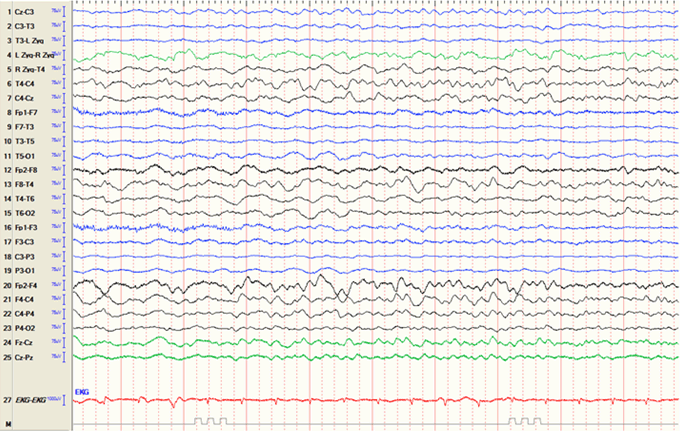
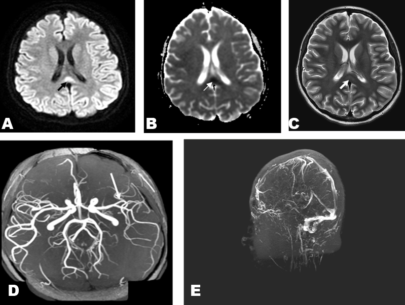
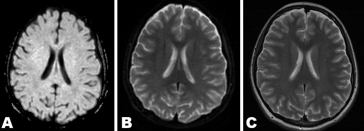
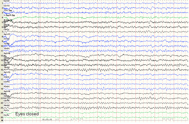
A 13-year-old right-handed Caucasian girl with no prior significant medical history was admitted for abdominal pain, fever, vomiting and bloody diarrhea for one week. She was being managed for HUS with hemodialysis and subsequently with plasma exchange (PLEX). She had positive stool test for Shiga toxin 2. Following her seventh cycle of PLEX, she reported to have a generalized tonic clonic seizure. On examination the patient was drowsy with a persistent left gaze deviation and right sided hemiplegia. She found to have diffuse hyperreflexia and non-sustained ankle clonus. She was afebrile with a temperature of 36.7°C (98°F) and normotensive with a blood pressure of 117/77 mmHg. She received 2 doses (2 mg) of intravenous lorazepam and loaded with 20 mg/kg of intravenous phenytoin. She was subsequently started on 100 mg three times daily dose of phenytoin. The patient was intubated due to respiratory insufficiency. She remained normotensive throughout this period. Long-term video EEG recording to evaluate for non-convulsive status revealed a diffusely slow background with significantly attenuated left hemispheric activity. Seizure like activity and epileptiform discharges were not seen (Figure 1). Brain magnetic resonance imaging (MRI) scan demonstrated diffusion restriction in the SCC with low apparent diffusion coefficient (ADC) correlates (Figure 2A–B). Axial FLAIR sequences revealed subtle hyperintensity in the SCC (Figure 2C). Magnetic resonance angiography (MRA) of the head revealed attenuation in the left middle cerebral artery (MCA) territory (Figure 2D). Magnetic resonance venogram (MRV) of the head was unremarkable (Figure 2E). Spinal fluid analysis indicated normal white blood cell count of 5x10³ cells/mm3 (36% neutrophils, 48% lymphocytes, 16% monocytes), a red blood cell count of 351 million cells/mm3, slightly increased protein of 68 mg/dl, and cerebrospinal fluid glucose of 81 mg/dl. Cerebrospinal fluid (CSF) gram stain and cultures were normal. Cerebrospinal fluid herpes simplex virus (HSV), varicella zoster virus (VZV), Epstein–Barr virus (EBV), cytomegalovirus (CMV), toxoplasma and west Nile virus were negative. The patient’s blood, urine and respiratory cultures were all negative. She had normal white blood cell count with normal comprehensive metabolic panel. Despite anticonvulsive therapy our patient continued to have left-gaze preference and right hemiparesis, which persisted for three days. It also became evident that despite being intubated she appeared to have a receptive aphasia. Given the MRA showed what appeared to be vasospasm of the left MCA it was elected to give a trial of intravenous MgSO4. She received 3 g (15 mg/kg) of MgSO4 in divided doses with significant improvement of her symptoms within three hours and complete resolution of her gaze preference, aphasia and hemiparesis within 24 hours. Twelve weeks after discharge from the inpatient setting her neurological examination, including cognitive and motor functioning, were completely normal. Repeat MRI scan of brain six weeks after onset of symptoms revealed complete resolution of the signal abnormalities in the SCC on DWI, ADC and axial FLAIR sequences (Figure 3A–C). Repeat long-term EEG recording eight weeks after her initial seizure was also normal (Figure 4). |
| ||||||
|
| ||||||
|
| ||||||
| ||||||
|
DISCUSSION
| ||||||
|
Neurological manifestation of HUS is considered to be multifactorial including hypertension, microangiopathy and metabolic derangements in the form of electrolyte imbalances secondary to dehydration and renal insufficiency. Most of the studies describe the neurological findings due to verotoxin induced damage to the endothelium of small vessels in the brain causing bleeding, infarction or cerebral edema due to microvascular changes which can be seen on neuroimaging [3][4]. These changes lead to secondary immune mediated cytokine release injury to the neuron and glial cells. Neurons and glia lack receptors for the verotoxin. Therefore, verotoxin is not directly neurotoxic [5]. Based on the animal studies Shiga toxin II is more neurotropic compared to Shiga toxin I [6] . This finding is also observed in children with HUS with serious neurological complications seen in patients with Shiga toxin II. The exact reason for comparatively increased virulence of Shiga toxin II is not known. However, it is speculated that since both Shiga toxin I and II compete for gut absorption through the same receptor so the absence of Shiga toxin I might increase the absorption of Shiga toxin II [6] . Our patient’s presentation with a seizure and focal neurological findings of left gaze deviation and right hemiparesis localizes to the left MCA territory which was evident as a possible left MCA vasospasm on MRA head, which correlated with her EEG findings of left hemispheric attenuation. The diffusion restricting lesions within SCC could be related to the disturbances in the intracerebral homeostasis in the absence of apparent metabolic derangements which possibly improved with the administration of intravenous MgSO4. Therapeutic role of MgSO4 has been extensively studied in cardiovascular and obstetrical practices. It is being used in the treatment of eclampsia and prevention of eclamptic seizures for many years. It is also used in the prophylactic treatment of angiogenic vasospasm in aneurismal subarachnoid hemorrhage. Magnetic sulfate is an NMDA receptor antagonist. It blocks the calcium influx through the voltage gated calcium channels and NMDA receptors; preventing smooth muscle contraction and neuronal damage [7] . It has been implicated that influx of calcium ions through NMDA receptor can lead to cerebral ischemic injury which can be reversible in the early stages of the disease pathogenesis [8]. Improvement of neurological symptoms in our patient after treatment with intravenous MgSO4 is a novel finding which has not been reported before in HUS. The left MCA vasospasm seen on MRA of head in our patient represents possibly early spectrum of the disease course before permanent cerebral ischemia or intracerebral hemorrhage. These changes responded to the treatment with MgSO4. We speculate that MgSO4 is not only involved in restoring the biochemical homeostasis and prevention of calcium influx through NMDA receptors but may also exert the NMDA modulating anti-inflammatory effect. This phenomenon can enhance the vasodilatation of the vessels as well as diminish the degree of immune mediated pathological processes leading to irreversible brain damage and irritation of the brain tissue causing seizures. Another interesting neuroimaging finding seen in our patient is reversible lesion in the SCC. This lesion demonstrated diffusion restriction and low ADC value on initial brain MRI scan represents cytotoxic edema. Reversible splenial lesion syndrome (RESLES) has been described in the literature and could be seen in variety of conditions including infections, seizures, metabolic disorders, malnutrition, high altitude cerebral edema and antiepileptic drug (AED) withdrawal [9]. Reversible splenial lesions are not commonly reported in patients with HUS. In our patient, this could represent an ongoing seizure activity or sequel of an underlying metabolic derangement causing deregulation of the cellular fluid. There are no clear guidelines for the treatment of neurological sequel of HUS. Gitaux et.al. suggested the use of monoclonal antibodies against the terminal complement pathway to treat the early signs of neurological involvement and prevent further complications in HUS. This study was based on the fact that Shiga toxin II seems to exert its effects through direct activation of the complement system [10]. Other treatments used for cerebral vasospasm include calcium channel blockers. The Magnesium sulfate induced vasodilatation and immune modulation can make it a reasonable treatment option in the early stages of vasospasms to stop the immune mediated pathological sequel as discussed above. | ||||||
|
CONCLUSION
| ||||||
|
Magnesium sulfate (MgSO4) plays an important role in restoring the biochemical homeostasis and prevention of calcium influx through N-methyl aspartate (NMDA) receptors. It may also exert the NMDA modulating anti-inflammatory effect which could potentially help preventing the reversible neuropathology associated with hemolytic uremic syndrome. | ||||||
|
REFERENCES
| ||||||
| ||||||
|
[HTML Abstract]
[PDF Full Text]
|
|
Author Contributions
Anza B. Memon – Substantial contributions to conception and design, Acquisition of data, Analysis and interpretation of data, Drafting the article, Revising it critically for important intellectual content, Final approval of the version to be published Salman Rashid – Substantial contributions to conception and design, Analysis and interpretation of data, Revising it critically for important intellectual content, Final approval of the version to be published Mitchel T. Williams – Substantial contributions to conception and design, Analysis and interpretation of data, Revising it critically for important intellectual content, Final approval of the version to be published |
|
Guarantor
The corresponding author is the guarantor of submission. |
|
Source of support
None |
|
Conflict of interest
Authors declare no conflict of interest. |
|
Copyright
© 2017 Anza B. Memon et al. This article is distributed under the terms of Creative Commons Attribution License which permits unrestricted use, distribution and reproduction in any medium provided the original author(s) and original publisher are properly credited. Please see the copyright policy on the journal website for more information. |
|
|
|
About the Authors
| |||||||||
| |||||||||
|
|