| |
|
|
|
Case Report
| ||||||
| Hypopharyngeal non-penetrating steel shrapnel foreign body: A case report of unusual route of impaction | ||||||
| Ahmad Nasrat Al-juboori1, Abdalla Mirghani Hamid2 | ||||||
|
1FICMS, EBE ORL-HNS, ENT Specialist, department of Surgery, Al Wakra Hospital, Hamad Medical Corporation, Doha, Qatar
2FRCS, ENT Senior Consultant, Al Wakra Hospital, Hamad Medical Corporation, Qatar | ||||||
| ||||||
|
[HTML Abstract]
[PDF Full Text]
[Print This Article]
[Similar article in Pumed] [Similar article in Google Scholar] 
|
| How to cite this article |
| Al-juboori AN, Hamid AM. Hypopharyngeal non-penetrating steel shrapnel foreign body: A case report of unusual route of impaction. Int J Case Rep Images 2017;8(5:313–316. |
|
ABSTRACT
| ||||||
|
Introduction: Foreign body injury is one of the most commonly encountered otorhinolaryngologic emergencies. The diagnosis and management of foreign bodies have mainly been based on the type and location of the foreign body. The workplace is a significant contributor to fatal and non-fatal injuries worldwide and an insufficiently appreciated contributor to the total burden of health care costs. Steel workers sustain a higher occupational hazard of penetrating injuries anywhere in the body, including the head and neck. However, we found no reports in literature about non-penetrating shrapnel foreign body injuries, particularly in the upper aero-digestive tract. Case series: A steelworker presented to the emergency department in Al Wakra hospital with a history of non-penetrating steel foreign body impaction in the throat which had been visualized by GlideScope and removed successfully with the assistance of Macintosh laryngoscope without complications. The purpose of this presentation is to highlight the unusual route taken by a shrapnel non-penetrating foreign body, through the open mouth to the hypopharynx. Conclusion:We concluded that this report could be regarded as the first case report of a non-penetrating steel foreign body with an unusual per oral route of impaction in the hypopharynx. It, also, highlighted its visualization and a comparison between two techniques of its extraction. Keywords: Foreign body throat, Glide video laryngoscope, Hypopharynx | ||||||
|
INTRODUCTION
| ||||||
|
Foreign body ingestion is one of the most commonly encountered otorhinolaryngologic disorders, often requiring urgent decision making and management. In particular, it has been reported that children younger than three years exhibit the greatest risk of foreign body swallowing [1][2]. In adults, foreign body ingestion occurs commonly, the majority of them pass spontaneously but some of them will impact at the hypopharyngeal level [3] . The result may be severe due to possible ulceration or even perforation with consequent life-threatening complications [4][5]. Many studies of foreign body injuries have focused on the case of young patients [2] [6]. However, it is also important to determine the frequency and characteristics of foreign body injuries in other age groups in addition to young children. Previous studies have demonstrated that some fatal cases occur in elderly patients due to foreign body asphyxia [7]. The diagnosis and management of foreign bodies have mainly been based on the type and location of the foreign body. Therefore, clinical information on the type and location of foreign bodies can expedite the management of these patients [7] . The workplace is a significant contributor to fatal and non-fatal injuries worldwide and an insufficiently appreciated contributor to the total burden of health care costs, like the occupational hazards of steel workers who manifest with penetrating injuries anywhere in the body[8]. However, there are no reported non-penetrating injuries of steel worker foreign body injuries particularly in the upper aerodigestive tract. The aim of the presentation of this case report is to highlight the route of a non-penetrating steel foreign body in the hypopharynx in a steelworker, and probably this could be the first reported case of this unusual route of entry. | ||||||
|
CASE REPORT
| ||||||
|
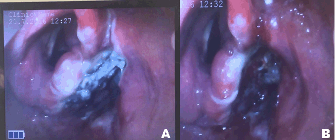
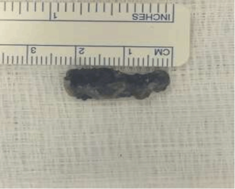
A 25-year-old steelworker presented to the emergency department in Al Wakra hospital with a history of foreign body impaction in the throat during work. When one of his colleagues was hammering steel nearby him, he felt something entering through his mouth, when it was open and getting inserted inside the throat. He was complaining of odynophagia, dysphagia with progressive change of voice. On examination, the patient was conscious, afebrile, not in respiratory distress and hemodynamically stable. Complete ear, nose and throat examination was normal, apart from a small wound at the tip of the tongue. Neck examination showed no evidence of external wounds or subcutaneous surgical emphysema but there was mild tenderness over the right side of the neck. flexible nasolaryngoscopy done under local anesthesia showed impacted foreign body (metal) on the right side of the supraglottic area with edema of the surrounding area of the epiglottis, aryepiglottic fold, arytenoids and part of the vocal fold on the right side. Plain X-ray of the neck, anteroposterior and lateral views, showed evidence of a metallic radio-opaque foreign body at the level of the fourth cervical vertebra directed towards the right side (Figure 1). The patient was prepared for examination under general anesthesia and the removal of the foreign body with the assistance of rigid scopes. Examination under general anesthesia in the supine position with full sedation and assistance of a GlideScope, revealed a clear video picture of the metallic foreign body in the right pyriform sinus (Figure 2A). Trial of removal through the GlideScope guidance failed, unfortunately, because of difficulties in directing the instrument towards the metallic foreign body. Then with the use of Macintosh laryngoscope the metallic foreign body was removed by forceps (Figure 3). Re-examination of the site of the foreign body was done again with the GlideScope and suction to the site of the foreign body to ensure there was no perforation (Figure 2B). In the postoperative period, the patient was kept for continuous intravenous fluids for the next 24 hours, with frequent checking of the neck for possible emphysema, then fluid diet was started for the next few hours and then the patient was discharged in good condition on the second postoperative day after psychiatric assessment to make sure that he is of normal mentality. | ||||||
| ||||||
| ||||||
| ||||||
|
DISCUSSION
| ||||||
|
Foreign body ingestion is more common in adults with mental developmental delay, psychiatric and neurological disorders or intoxication and in patients with dentures or dental bridges because of the decrease in tactile sensation during swallowing. The most commonly ingested foreign bodies are fish and chicken bones and there is an apparent predominance of certain types in specific groups of patients [9] e.g., coins and toys in children, razor blades and cutlery in prisoners [10]. Our case report highlights the peculiar route taken by the shrapnel foreign body, through the open oral cavity to the hypopharynx, a route we think have not been reported before. Also, it appears that the foreign body has taken a curved projectile rather than a straight pathway. This is suggested by the fact that no injuries were found in the oral cavity or oropharynx, apart from a small laceration at the tip of the tongue. Medline search did not report this kind of foreign body route of entry, so we consider this is the first reported case with such description. GlideScopes represent a recent advancement over the Macintosh laryngoscope. It has increased endotracheal intubation success rate, and it is recommended mainly in the management of potential difficult airways in several patient populations [11]. The use of these devices to remove foreign bodies has been reported previously only in two adult patients affected by a non-impacted partial denture in the hypopharynx [12]. While desirable, it is not reported so far in the pediatric population. Je et al., in a cadaver study, compared Macintosh laryngoscope and the GlideScope for extracting hypopharyngeal foreign bodies. He stated that it is not possible to conclude that videoscopes are inferior to the Macintosh laryngoscope for foreign body extractions, and that the GlideScope might be necessary to aid the foreign body extraction in emergency situations [13]. In our case report, the GlideScope provided good indirect visualization and localization with magnification of the foreign body but it did not provide good access for removal. On the other hand, the Macintosh provided a direct visualization of the foreign body and easy removal and extraction. | ||||||
|
CONCLUSION
| ||||||
|
From previous descriptions, we concluded that this report could be regarded as the first case report of a non-penetrating steel foreign body with an unusual per oral route of impaction in the hypopharynx. It, also, highlighted its visualization and a comparison between two techniques of its extraction. | ||||||
|
REFERENCES
| ||||||
| ||||||
|
[HTML Abstract]
[PDF Full Text]
|
|
Acknowledgements
We would like to thank all theater staff in AlWakra hospital for their help and support. |
|
Author Contributions
Ahmad Nasrat Al-juboori – Substantial contribution to conception and design, Acquisition of data, Drafting the article, revising it critically for important intellectual content; Final approval of the version to be published Abdalla Mirghani Hamid – Substantial contribution to conception and design, Acquisition of data, Drafting the article, Revising it critically for important intellectual content, Final approval of the version to be published |
|
Guarantor
The corresponding author is the guarantor of submission. |
|
Source of support
None |
|
Conflict of interest
Authors declare no conflict of interest. |
|
Copyright
© 2017 Ahmad Nasrat Al-juboori et al. This article is distributed under the terms of Creative Commons Attribution License which permits unrestricted use, distribution and reproduction in any medium provided the original author(s) and original publisher are properly credited. Please see the copyright policy on the journal website for more information. |
|
|