| |
|
|
|
Case Report
| ||||||
| Leiomyoma of hard palate: A rare case report | ||||||
| G. Siva Prasad Reddy1, B. Jagannadha Prasad2, A. Bhargavi Krishna3, P.S.S.Tejaswini4, J. Laxmi Sravya4, S. Sushma4 E. Padmini4 | ||||||
|
1MDS, Professor, Department of Oral and Maxillofacial Surgery, Panineeya Mahavidyalaya Institute of Dental Sciences and Research Centre, Road No 3, Kamalanagar, Dilsukhnagar, Hyderabad, Telangana, India
3MS DLO, Associate Professor, Department of Otorhinolaryngology & Head and Neck Surgery, Mediciti Institute of Medical Sciences, Medchal mandal, Ghanpur, Hyderabad, Telangana, India
3MDS, Professor, Department of Oral and Maxillofacial Pathology, Panineeya Mahavidyalaya Institute of Dental Sciences and Research Centre, Road No 3 Kamalanagar, Dilsukhnagar, Hyderabad, Telangana, India
4BDS, Post Graduate Student, Department of Oral and Maxillofacial Surgery, Panineeya Mahavidyalaya Institute of Dental Sciences and Research Centre, Road No 3, Kamala Nagar, Dilsukhnagar, Hyderabad, Telangana, India.
| ||||||
| ||||||
|
[HTML Abstract]
[PDF Full Text]
[Print This Article]
[Similar article in Pumed] [Similar article in Google Scholar] |
| How to cite this article |
| Reddy GSP, Prasad BJ, Krishna AB, Tejaswini PSS, Sravya L, Sushma S, Padmini E. Leiomyoma of hard palate: A rare case report. Int J Case Rep Images 2017;8():270–274. |
|
Abstract
| ||||||
|
Introduction: Leiomyomas are benign, soft tissue tumors, arising from the smooth muscle. They usually affect the muscular layer of the gut and body of the uterus. Oral leiomyomas are extremely rare and are thought to arise from the smooth muscle wall of blood vessels. They present as slow growing, well-circumscribed, painless swellings accounting for only 0.42% of soft tissue neoplasms [1] of the oral cavity.
Keywords: Leiomyoma, Oral leiomyoma, Smooth muscle tumor, Hard palate tumor | ||||||
|
Introduction
| ||||||
|
Leiomyomas are benign soft tissue tumors arising from the smooth muscle due to nodular proliferation of muscle cells. Primary leiomyomas of head and neck account for 12% of all leiomyomas [2]. It is more commonly seen in the uterine myometrium (95%), skin (3%), gastrointestinal tract (1.5%) and less than 1% is seen in the head and neck region [3] . It rarely affects the oral cavity due to absence of smooth muscle except in the wall of blood vessels. Oral leiomyomas are rare (0.06%) and they mostly occur on the lips, tongue, palate and cheek region. Usually, they present as painless, slow growing masses. Occasionally they may cause pain, mobility of adjacent teeth and difficulty in chewing. Due to its unspecific clinical presentation diagnosis is made after histopathological study. Immunohistochemical studies offer a more precise diagnosis in these lesions. Surgical resection of the lesion is the treatment of choice. In this article, we report a case of oral leiomyoma of hard palate in a 14-year-old boy who reported to our department with a chief complaint of painless swelling in the palate. | ||||||
|
Case Report
| ||||||
|
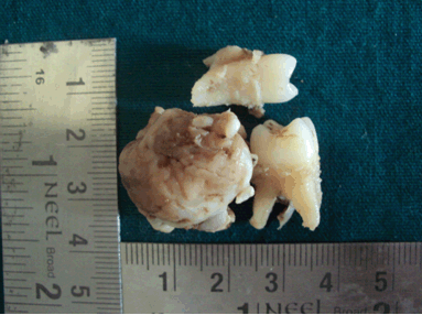
A 14-year-old boy presented to our department with a chief complaint of slow-growing mass since six months in the left hard palate. It was initially small when noticed and steadily progressed to the present size (2x2 cm approx). It was a painless swelling and interfered during chewing food. The patient was otherwise healthy, with no systemic abnormalities and no deleterious or para-functional habits. Clinical examination revealed a sessile, well-circumscribed swelling in the left palate region extending from maxillary first premolar to maxillary first molar (Figure 1). Swelling extended onto the occlusal and buccal surfaces of maxillary second premolar. It was non-tender on palpation and soft in consistency. Overlying mucosa was reddish pink in appearance. There was slight cortical plate expansion in the region of maxillary second premolar. No cortical perforation was noted. Lymph nodes were not palpable. We have advised fine needle aspiration biopsy and CBCT of upper left maxillary region. Aspiration results were negative, thus ruling out vascular and cystic lesions. CBCT revealed alveolar bone loss in relation to left maxillary second premolar and left maxillary first molar (Figure 2). The case was planned for excisional biopsy under general anesthesia owing to its size and anatomical location. Complete resection of the tumor mass along with the extraction of upper left second premolar and first molar was done under general anesthesia (Figure 3). The resected specimen (Figure 4) was sent for histopathological examination, which suggested the presence of numerous spindle shaped cells (Figure 5). with cigar shaped nucleus and endothelial cells in a sparse stroma without any atypia. To arrive at a more definitive diagnosis, we further subjected the specimen to immunohistochemical studies. The tumor exhibited strong positivity for SMA (Figure 6) and vimentin (Figure 7), thus confirming leiomyoma. No recurrence was noted during the four month follow-up period. | ||||||
| ||||||
| ||||||
| ||||||
| ||||||
| ||||||
| ||||||
| ||||||
|
Discussion
| ||||||
|
Leiomyomas are benign neoplasms that arise from smooth muscle. Virchow [4] in 1854 first described this tumor. Although these are rare tumors of the head and neck region, when they occur, they are commonly seen in the oral cavity, nasal cavity and larynx [5]. The hereditary form causing multiple leiomyoomas was noted by Kloepfer et al. [6] in 1958. Smooth muscle tumors are relatively rare in the oral cavity, accounting for only 0.42% of soft tissue tumors because of the paucity of smooth muscle in this region [7] . The only source of smooth muscle in the oral cavity is tunica media as suggested by Scout [8], or the ductus lingualis or circumvallate papillae as suggested by Glass [9]. Brooks et al. reported that, the most frequently affected site was the lip (43.6%) followed by the palate (21.1%), buccal mucosa and tongue (each 9.2%), mandible (8.3% ) and buccal and labial sulcus[10]. The WHO classified leiomyomas into three groups angioleiomyoma (74%), solid leiomyoma (25%) and epitheloid leiomyoma (1%). Leiomyoma differs from angioleiomyoma in the degree of angiogenesis [11]. In 1884, the first case of oral leiomyoma was reported by Blanc [12] in a 33-year-old male who presented with a large-tumor at the base of the tongue. Oral leiomyomas are benign neoplasms that present as small, solitary, asymptomatic nodular mass. Highest prevalence is seen in the 4th and 5th decades of life with slight male predilection [13] (1.43:1). However, few reports suggest slight female predominance of leiomyomas in the head and neck region and the authors attribute it to hormonal variation, i.e., progesterone receptor positive and estrogen receptor negative on immunochemical studies [14][15]. Although most of these lesions are asymptomatic, few authors reported symptomatic lesions that are associated with pain, tooth mobility, difficulty in chewing and swallowing. Pain when occurs can be due to local ischemia causing intra-tumoral vasoconstriction or compression of a somatic nerve by the tumor mass [10]. The average size of these tumors as reported in literature is 1–2 cm with less than 1 year duration. The color of the lesion depends on depth and vascularity. Clinically, it is difficult to differentiate a leiomyoma from other mesenchymal tumors or its malignant counterpart. Hence, the final diagnosis of leiomyoma is mainly determined by histopathological examination. Leiomyomas are well encapsulated lesions, typically composed of numerous spindle shaped/ fusiform mesenchymal cells arranged in whorls or strands. The nucleus is typically elongated and cigar shaped with eosinophilic cytoplasm on Hematoxylin and Eosin staining. Endothelial cells are seen lining the vascular channels. Unlike other mesenchymal tumors, leiomyomas lack dense fibrous stroma in between the individual mesenchymal cells. As the tumor cells mimic fusiform cells, simple Hematoxylin and Eosin staining cannot differentiate between leiomyomas and other spindle cell tumors. Special stains to identify collagen and muscle cells, such as Von Gieson stain, Masson trichromic acid stain, Mallory’s phosphotungstic acid-hematoxylin (PTAH) stain can be used [16]. Von Gieson stain is recommended for muscle. Masson trichromic acid stains the cytoplasmic elements of smooth muscle cells red and collagen and fibroblasts blue or green. Myofibrils are stained by Mallory PTAH stain [17]. Additionally, immunohistochemical studies can be used for a more precise diagnosis. Specific monoclonal antibodies for actin (smooth muscle marker) confirm leiomyoma. Smooth muscle actin that corresponds to the alpha fraction of actin chain is an immuno-marker for smooth muscle but it can have immune reaction to skeletal muscle. S100, vimentin, desmin, antiCD-34 antibody are the other immunomarkers. Vimentin is a structural protein of cytoplasmic elements of mesenchymal cells whereas desmin is a type III intermediate filament near the Z line in sarcomere [18]. CD-34 is a transmembrane protein, expressed by the vascular endothelium and endothelial cells exhibit strong immunoreactivity against antiCD-34 antibody [19]. Leiomyoma should be carefully differentiated from its malignant counterpart i.e., leiomyosarcoma. Clinically, the presence of ulceration may be indicative of malignancy. Cotran [20] et al. described few histological features in uterine tumors, which were suggestive of malignancy like presence of more than 10 mitoses per 40x high magnification field, with or without cellular atypia or 5–10 mitoses per x10 magnification field with atypia . Tumors with 1–4 mitoses per x10 magnification, necrotic areas and atypias are best considered potentially malignant. Presence of fewer than two mitoses in x10 magnification indicates good prognosis. Molecular markers like PCNA, bcl-2, CDK-4, P53, MDM 2 indicate malignancy [21]. In our case, the resected tumor showed numerous spindle cells on Hematoxylin and Eosin staining arranged in parallel bundles with endothelial lining and no atypia. Hence, to differentiate it from other spindle cell tumors, immunohistochemical staining has been done. The tumor cells stained positive for vimentin and smooth muscle actin, thus confirming the diagnosis of leiomyoma. The treatment of choice is local resection with adequate safety margins. Leiomyomas are tumors of vascular origin, but bleeding after resection is not routinely seen. Benign smooth muscle tumors rarely relapse. Brooks et al. reported relapse of two cases , two weeks and nine months postoperatively following resection of hard palate leiomyomas [9]. | ||||||
|
Conclusion
| ||||||
|
Leiomyomas are benign tumors of smooth muscle origin. Oral leiomyomas are relatively rare. Definitive diagnosis is made after histological and immunohistochemical confirmation. Surgical resection of the tumor with adequate safety margins is the preferred treatment. Recurrence is rarely seen following excision in these tumors. | ||||||
|
References
| ||||||
| ||||||
|
[HTML Abstract]
[PDF Full Text]
|
|
Author Contributions
G. Siva Prasad Reddy – Substantial contributions to conception and design, Acquisition of data, Analysis and interpretation of data, Drafting the article, Revising it critically for important intellectual content, Final approval of the version to be published B. Jagannadha Prasad – Substantial contributions to conception and design, Acquisition of data, Analysis and interpretation of data, Drafting the article, Revising it critically for important intellectual content, Final approval of the version to be published A. Bhargavi Krishna – Substantial contributions to conception and design, Acquisition of data, Analysis and interpretation of data, Drafting the article, Revising it critically for important intellectual content, Final approval of the version to be published P.S.S. Tejaswini – Substantial contributions to conception and design, Acquisition of data, Analysis and interpretation of data, Drafting the article, Revising it critically for important intellectual content, Final approval of the version to be published J. Laxmi Sravya – Substantial contributions to conception and design, Acquisition of data, Analysis and interpretation of data, Drafting the article, Revising it critically for important intellectual content, Final approval of the version to be published S. Sushma – Substantial contributions to conception and design, Acquisition of data, Analysis and interpretation of data, Drafting the article, Revising it critically for important intellectual content, Final approval of the version to be published E. Padmini – Substantial contributions to conception and design, Acquisition of data, Analysis and interpretation of data, Drafting the article, Revising it critically for important intellectual content, Final approval of the version to be published |
|
Guarantor
The corresponding author is the guarantor of submission. |
|
Source of support
None |
|
Conflict of interest
Authors declare no conflict of interest. |
|
Copyright
© 2017 G. Siva Prasad Reddy et al. This article is distributed under the terms of Creative Commons Attribution License which permits unrestricted use, distribution and reproduction in any medium provided the original author(s) and original publisher are properly credited. Please see the copyright policy on the journal website for more information. |
|
|