  |

|
 |
|
Case Report
| ||||||
| Scrotal swelling in a patient with paroxysmal supraventricular tachycardia | ||||||
| Riccardo Bentivegna1, Domenico Nobile1, Giuseppina Novo2, Salvatore Novo3 | ||||||
|
1MD, Doctor, cardiology, University Hospital “Paolo Giaccone”, Palermo, Italy 2MD, Professor of Cardiovascular Diseases, cardiology University Hospital “Paolo Giaccone”, Palermo, Italy 3MD, Professor of Cardiovascular Diseases – FESC, cardiology, University Hospital “Paolo Giaccone”, Palermo, Italy | ||||||
| ||||||
|
[HTML Abstract]
[PDF Full Text]
[Print This Article]
[Similar article in Pumed] [Similar article in Google Scholar] 
|


| How to cite this article |
| Bentivegna R, Nobile D, Novo G, Novo S. Scrotal swelling in a patient with paroxysmal supraventricular tachycardia. Int J Case Rep Images 2017;8(4):257–260. |
|
Abstract
| ||||||
|
We present a case of a patient with paroxysmal supraventricular tachycardia and a history of chronic ischemic disease evolved into dilated cardiomyopathy. During hospitalization, the occurrence of arrhythmic crisis, our patient felt sporadic palpitations associated with dyspnea, distended jugular veins and scrotal swelling, quite unexpected clinical sign that caught our attention. Echocardiography showed dilated left ventricle, severely reduced systolic function, dilated right-sided, dilated inferior vena cava with reduced respiratory excursions. Echography of both testicles showed bilateral expansion of pampiniform plexus with reflux to the functional maneuvers, such as varicocele of grade III–IV. Afterwards coronary angiography, the placement of a metallic stent in left anterior descending artery improved coronary circulation with the net reduction of dyspnea. In later episodes of supraventricular tachycardia the only symptom that the patient felt was mild palpitation associated with scrotal swelling and distended jugular veins. The increased heart rate led to an exacerbation of the underlying pulmonary hypertension resulting in venous stasis upstream the pulmonary capillaries. Restoring sinus rhythm, the heart performs its normal pump function and the pressure within the pulmonary capillaries was decreased, so on physical examination turgor of the jugular veins and scrotal size were strongly reduced. Keywords: Paroxysmal supraventricular tachycardia, Pulmonary hypertension, Scrotal swelling, Varicocele | ||||||
|
Introduction
| ||||||
|
Paroxysmal supraventricular tachycardia (PSVT) is an acronym that includes all of the arrhythmias that originate above the ventricles and that manifest paroxysmally. The causes of these arrhythmias may be different. The most common mechanism is the presence within the heart of a ‘short circuit’ due to the presence of an anatomical or functional so-called accessory pathway. The most common forms of PSVT are atrioventricular nodal reentrant tachycardia (AVNRT) and atrioventricular reentry tachycardia (AVRT). The atrioventricular nodal reentrant tachycardia is characterized by the presence of this reentry circuit in the atrioventricular node [1]. The reentry functional circuit is constituted by two parts with different impulse conduction velocity, and tachycardia is generated when the impulse can circulate inside the reentry circuit [2]. The atrioventricular nodal reentrant tachycardia is a benign arrhythmia in most cases, whose main symptoms are palpitations and shortness of breath (although not always present) [3] . | ||||||
|
Case Report
| ||||||
|
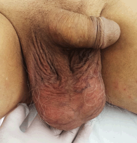
A 66-year-old Italian male, with a history of high blood pressure and chronic ischemic heart disease (two prior PCI in the circumflex coronary artery) evolved into dilated cardiomyopathy, reaches the emergency room of our hospital because of onset of worsening dyspnea. On admission, dyspnea was reduced, blood pressure was 130/70 mmHg, arterial oxygen saturation was 99%, blood test demonstrated normal levels of cardiac troponin I (82 ng/ml), elevated creatinine (1.46 mg/dl) with eGFR 51 ml/min/m2, elevated white blood cell count (12 x106/L). Chest X-ray showed no ilo-pleural-parenchymal lesions in active phase. During the next few hours, the patient becomes progressively dyspneic and asthenic and continuous electrocardiography monitoring showed two episodes of PSVT with heart rate of 160 bpm, both regressed with restoration of sinus rhythm after carotid sinus massage (Figure 1) (Figure 2). The patient was, therefore, transferred to our cardiology department for the continuation of the diagnostic and therapeutic iter. The patient appeared confused and with loss of memory. Physical examination revealed heart tones in rhythmic succession, systolic murmur 2–3/6L more audible on the mitral outbreak, slightly reduced breath sounds in all lung fields and fine crackles to the basic fields. 12-lead electrocardiography showed: sinus rhythm, previous necrosis in the inferior leads, repolarization abnormalities in the inferior and posterolateral leads. It was set aspirin therapy, omeprazole, fondaparinux, furosemide, ciprofloxacin, amiodarone, saline 0.9% NaCl infusion at 30 ml/h. During hospitalization, the occurrence of arrhythmic crisis, our patient felt sporadic palpitations and dyspnea associated with the appearance of distended jugular veins and scrotal swelling (Figure 3), quite unexpected clinical sign that caught our attention, on physical examination. Considering the clinical-anamnestic context it was performed coronary angiography that showed a critical stenosis of 70% left anterior descending coronary artery (LAD) and good patency of the prior stents in circumflex coronary artery. It was therefore performed a percutaneous transluminal coronary angioplasty (PTCA) with metal stent placement in LAD. Improving cardiac perfusion, ongoing of arrhythmia, our patient does not feel the symptoms described above. However, at the following episodes of PSVT, on physical examination persisted scrotal swelling (already observed before the angioplasty procedure) and the appearance of distended jugular veins. Echocardiography showed dilated left ventricle (VTD 160 ml - DTD 65 mm) with wall thickness increased; severely reduced systolic function (EF 28%); akinesia of the lower and posterolateral wall, hypokinesis of the remaining segments; severely dilated left atrium (150 ml); regular size of the aortic bulb and ascending aorta ectasia (39 mm); dilated right-sided with reduced TAPSE; dilated inferior vena cava with reduced respiratory excursions; thickened mitral leaflets with symmetrical tethering; sclerosis of the semilunar aortic valves; moderate mitral regurgitation with restrictive pattern, mild aortic regurgitation, moderate tricuspid regurgitation. Echography of both testicles performed through high-frequency probe (12 MHz) showed normal morpho-volumetric and eco-structural appearance of both didymus and epididymis; regular intraparenchymal vascularization to the color Doppler evaluation; bilateral expansion of pampiniform plexus (maximum right vessel caliber 2.6 mm - maximum left vessel caliber 3.6 mm) with reflux to the functional maneuvers, such as varicocele of grade III-IV. Coexisted diffuse thickening of the subcutaneous soft tissues such as edematous imbibition (Figures 4A–B). | ||||||
| ||||||
| ||||||
| ||||||
| ||||||
|
Discussion
| ||||||
|
The veins that originate from pampiniform plexus, after the passage along the inguinal canal, join to form a single testicular vein, which opens on the right side into the inferior vena cava (has an high flow) at an acute angle; on the left side into the left renal vein (has low flow) at a right angle [4] These veins can become incontinent and dilate thereby preventing venous outflow of blood from the testicular veins to the inferior vena cava. This creates a condition of reflux and stasis of blood to the testicle. This is manifested in particular in the left testicle (95%) and rarely in the right testicle (5%) because of the different anatomical features between the two vascular pathways [5]. A complete physical examination in orthostatic position allows highlighting the expansion of veins during the Valsalva maneuver. Instrumental support investigations (ultrasound and testicular color Doppler of the spermatic vessels) allow to determine the extent of reflux and measure the diameters of the testicles [6] [7] . Current guidelines define pulmonary hypertension as a hemodynamic and pathophysiological condition characterized by an increase in mean pulmonary arterial pressure (PAPm) =25 mmHg at rest as assessed by right heart catheterization (RHC). Available data have shown that the normal PAPm at rest is 14±3 mmHg [8]. The secondary forms of pulmonary hypertension may result from [9]:
Symptoms of pulmonary hypertension are very unspecific. However, shortness of breath appears to be the most common initial symptom (60% of cases) and is present in 98% of patients at diagnosis. Other disorders, less frequent in the early stages, are often observed at diagnosis, and include fatigue, weakness (73% at diagnosis), chest pain (47%), fainting (41%), syncope (36%), lower limb edema (37%) and palpitations (33%)[10]. | ||||||
|
Conclusion
| ||||||
|
Scrotal swelling, quite unexpected clinical sign, in our case report was secondary to the patient’s pulmonary hypertension. It was present both as a result of cardiac causes (left ventricular failure and moderate mitral regurgitation), both as a result of pulmonary causes (history of COPD). The increased pressure developed in the pulmonary circulation, determines upstream blood stagnation. It was observed instrumentally on echocardiography with a dilated inferior vena cava and poor respiratory excursions, and clinically with distended jugular veins and the sudden and temporary scrotal swelling during the PSVT episode (was confirmed by ultrasound with the detection of varicocele grade IV and the high blood stasis). Restoring sinus rhythm, the pressure within the pulmonary capillaries decreased, so on physical examination turgor of the jugular veins and scrotal size were strongly reduced. | ||||||
|
References
| ||||||
| ||||||


|
[HTML Abstract]
[PDF Full Text]
|
|
Author Contributions
Riccardo Bentivegna – Substantial contributions to conception and design, Acquisition of data, Analysis and interpretation of data, Drafting the article, Revising it critically for important intellectual content, Final approval of the version to be published Domenico Nobile – Analysis and interpretation of data, Revising it critically for important intellectual content, Final approval of the version to be published Giuseppina Novo – Analysis and interpretation of data, Revising it critically for important intellectual content, Final approval of the version to be published Salvatore Novo – Analysis and interpretation of data, Revising it critically for important intellectual content, Final approval of the version to be published |
|
Guarantor
The corresponding author is the guarantor of submission. |
|
Source of support
None |
|
Conflict of interest
Authors declare no conflict of interest. |
|
Copyright
© 2017 Riccardo Bentivegna et al. This article is distributed under the terms of Creative Commons Attribution License which permits unrestricted use, distribution and reproduction in any medium provided the original author(s) and original publisher are properly credited. Please see the copyright policy on the journal website for more information. |
|
|