| |
|
|
|
Case Report
| ||||||
| Duplication cyst in the sigmoid colon mimicking a submucosal tumor: A case report | ||||||
| Pyong Wha Choi1, Mee Joo2 | ||||||
|
1MD, PhD, Department of Surgery, Ilsan Paik Hospital, Inje University College of Medicine, Goyang-si, Gyeonggi-do, South Korea 2MD, PhD, Department of Pathology, Ilsan Paik Hospital, Inje University College of Medicine, Goyang-si, Gyeonggi-do, South Korea | ||||||
| ||||||
|
[HTML Abstract]
[PDF Full Text]
[Print This Article]
[Similar article in Pumed] [Similar article in Google Scholar] |
| How to cite this article |
| Choi PW, Joo M. Duplication cyst in the sigmoid colon mimicking a submucosal tumor: A case report. Int J Case Rep Images 2017;8(4):248–251. |
|
Abstract
| ||||||
|
Introduction: A duplication cyst in the alimentary tract is a rare congenital anomaly that most often occurs in the small bowel and rarely occurs in the colon. Most cases in the small bowel are diagnosed at childhood due to complications or symptoms caused by the duplication cyst. However, duplication cysts in the colon are often asymptomatic, despite resulting in rare complications, such as obstruction, infection, and inflammation. Thus, duplication cysts in the colon can be detected incidentally through imaging studies or during a colonoscopy, causing them to possibly be misdiagnosed as a submucosal tumor of the colon.
Keywords: Colon, Cyst, Duplication | ||||||
|
Introduction
| ||||||
|
A duplication cyst of the alimentary tract is a rare congenital anomaly that is defined by its cystic structure that exhibits a common muscular wall and bloody supply with the nearby alimentary tract[1]. While these cysts can develop along the entirety of the gastrointestinal tract, the ileum is the most common site. Meanwhile, the development of a duplication cyst in the colon is rare [2][3] [4]. Duplication cysts can be detected in any age, but pediatric patients comprise most of the cases because of the symptoms caused by the cysts [2][3][4]. In adults, duplications cysts are often diagnosed by chance during an imaging study or colonoscopy. Herein, we present a case of a 64-year-old male with a duplication cyst in the sigmoid colon, which was preoperatively diagnosed as a large submucosal tumor. | ||||||
|
Case Report
| ||||||
|
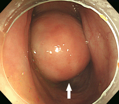
A 64-year-old male was referred to the department of surgery of Inje University, College of Medicine, Ilsan Paik Hospital for the treatment of a submucosal mass in the sigmoid colon. He had undergone a colonoscopy one week ago at a local clinic to screen for colorectal cancer and was referred to the department of gastroenterology for further evaluation and management. He had no past medical history and did not complain of any symptoms related to the mass. Upon admission, his vital signs were stable and his laboratory results were within normal limits, including those of tumor markers. The colonoscopy revealed a large round submucosal mass in the sigmoid colon. The mass was of a hard consistency when pressed with a scope forceps (Figure 1). Abdominopelvic computed tomography scan indicated an approximately 4 cm homogeneous enhancing mass in the sigmoid colon and there was no evidence of lymphadenopathy (Figure 2). With the presumed diagnosis of a submucosal tumor, such as gastrointestinal stromal tumor (GIST), laparoscopic anterior resection was performed. Subsequently, histologic examination was performed, which indicated that the mass was a duplication cyst of the sigmoid colon. The cyst was lined by colonic-type mucosa and had a thin muscular layer partly shared with the adjacent colonic wall (Figure 3). The postoperative course was uneventful, and the patient was discharged on postoperative day-7. | ||||||
| ||||||
| ||||||
|
| ||||||
|
Discussion
| ||||||
|
Gastrointestinal duplication is a rare congenital anomaly that is mostly diagnosed before the age of two years [3][4]. Although several theories exist regarding the development of gastrointestinal duplication, the etiology and pathogenesis have not been well established [2]. As the disease name implies, diagnosis is made on the basis of pathologic findings of a tubular or cystic structure that tends to be located on the mesenteric side of the bowel, shares a common muscular wall and blood supply with the bowel wall, and a separate mucosal lining [1]. Communication with the bowel lumen may or may not exist [5]. Although gastrointestinal duplication has been classified according to various pathologic criteria, it is simply divided into tubular or cystic in nature. Most gastrointestinal duplications are cystic structures and the ileum is the most common site [3][4]. Colonic involvement accounts for less than 20% of cases of gastrointestinal duplication. Of these colonic duplications, 40% are located in the cecum, with the sigmoid being rarely involved [4]. The clinical manifestation of duplication cysts is variable and range from non-specific abdominal pain to peritonitis. The location, size, communication with the bowel lumen, and presence of the heterotopic mucosa, such as ectopic gastric mucosa, are the main determinants for the development of symptoms, including perforation, obstruction, and bleeding [2][3][4][5][6] . Most gastrointestinal duplication cysts are detected during childhood due to complication or related symptoms [2][3] . Even though colonic duplication cyst may be a leading point resulting in colonic obstruction by intussusception, and infection, inflammation, and ulceration of the cyst may lead to the symptom and sign like diverticulitis and Crohn’s disease, colonic duplication cyst tends to remain asymptomatic [3][6] [7]. Thus, duplication cysts can be incidentally found in adults during colonoscopy or imaging studies for other reasons, such as that which occurred in our case. The diagnosis of a duplication cyst may be made with imaging studies, such as ultrasonography, computed tomography (CT) scan, magnetic resonance imaging (MRI) scan, and contrast study [2][3] [8][9]. Ultrasonography is the imaging modality of choice in childhood patients because it provides highly accurate images and avoids the risk of radiation [3][9]. However, its accuracy is operator dependent, and it may not be feasible in patients with complication. For cases in which Ultrasonography findings are equivocal or Ultrasonography is not feasible, a CT scan or MRI scan are useful modalities to establish the diagnosis given the nature, location, and extent of the lesion [2][8]. More recently, endoscopic ultrasonography (EUS) and EUS-guided fine needle aspiration have been applied for the diagnosis of colonic submucosal tumor. However, the role of EUS has not been established in the diagnosis of colonic duplication cyst. Contrast studies and colonoscopy are diagnostic if there is sufficient communication to allow contrast or a scope to pass between the true lumen of the colon and the cyst [2]. However, when there is no communication, diagnosis though contrast studies or colonoscopy is limited because the lesion can appear similar to a submucosal tumor. Thus, some authors have suggested that if the diagnosis is unclear, then laparoscopy may be used to confirm the diagnosis [3] . In the present case, although we performed laparoscopic surgery, the final diagnosis was made through pathologic examination because of the rarity of the case. Therefore, duplication of the colon may be considered one of the differential diagnoses in a patient presenting with cystic colonic mass. While the treatment of choice is not well established in asymptomatic patients, the treatment in symptomatic or diagnosed patients is surgical excision [4][6] . Surgical treatment options range from simple cystectomy to en block resection with the adjacent colon. Since colon cancer may develop in a duplication cyst, and since laparoscopic colon resection is feasible, then en block resection may be a reasonable treatment option in patients with a colonic duplication cyst. | ||||||
|
Conclusion
| ||||||
|
Although duplication cysts in the colon are rare, when a degenerative cystic colon mass is detected, a duplication cyst might be considered as one of the differential diagnoses. | ||||||
|
References
| ||||||
| ||||||
|
[HTML Abstract]
[PDF Full Text]
|
|
Author Contributions
Pyong Wha Choi – Substantial contributions to conception and design, Acquisition of data, Analysis and interpretation of data, Drafting the article, Revising it critically for important intellectual content, Final approval of the version to be published Mee Joo – Substantial contributions to conception and design, Acquisition of data, Analysis and interpretation of data, Final approval of the version to be published |
|
Guarantor
The corresponding author is the guarantor of submission. |
|
Source of support
None |
|
Conflict of interest
Authors declare no conflict of interest. |
|
Copyright
© 2017 Pyong Wha Choi et al. This article is distributed under the terms of Creative Commons Attribution License which permits unrestricted use, distribution and reproduction in any medium provided the original author(s) and original publisher are properly credited. Please see the copyright policy on the journal website for more information. |
|
|