| |
|
|
|
Case Report
| ||||||
| Umbilical metastasis as a primary presentation in carcinoma rectum: A case report | ||||||
| Syed Altaf1, Kapil Dev2, Jaiprakash Gurawalia2, Shiva Kumar2 | ||||||
|
1Associate Professor, Department of Surgical Oncology, Kidwai Memorial Institute of Oncology, Bangalore.
2Mch student, Department of Surgical Oncology, Kidwai Memorial Institute of Oncology, Bangalore, Karnataka, India. | ||||||
| ||||||
|
[HTML Abstract]
[PDF Full Text]
[Print This Article]
[Similar article in Pumed] [Similar article in Google Scholar] 
|
| How to cite this article |
| Altaf S, Dev K, Gurawalia J, Kumar S. Umbilical metastasis as a primary presentation in carcinoma rectum: A case report. Int J Case Rep Images 2017;8(3):201–204. |
|
Abstract
|
|
Introduction:
Sister Mary Joseph nodule refers to periumbilical metastatic lesion and is an indicator of advanced intra-abdominal malignancy. It can be smooth, non-ulcerated or ulcerated and necrotic mass with or without blood, mucinous, serous or purulent discharge. It can appear before the diagnosis of the primary lesion or during or after the definitive treatment.
Case Report: A 39-year-old man presented with a two-month history of painful swelling over the umbilicus. On workup, umbilical nodule was diagnosed as metastatic adenocarcinoma with a primary lesion in mid rectum. Conclusion: Sister Mary Joseph nodule is a thumbprint of disseminated and advanced disease that requires an aggressive combined treatment in every individual instance and bears poor prognosis. | |
|
Keywords:
Carcinoma rectum, Metastatic adenocarcinoma, Sister Mary Joseph nodule
| |
|
Introduction
| ||||||
|
Metastatic umbilical lesion also known as Sister Mary Joseph nodule (SMJN) is secondary to a primary malignancy of any viscera. So far reported cases of SMJN can be categorized as metastatic from gastrointestinal malignancies (35–65%), genitourinary tract (12–35%), unknown sites (15–30%) and those from the lung and breast (3–6%) [1]. Depending on the characteristics of the primary tumor inside, it can be fissured or ulcerated and secreting serous, mucinous, purulent or bloody discharge [1] [2]. Umbilical metastasis from intra-abdominal visceral malignancies is a form of SMJN. It can be a presenting feature of undiagnosed malignancy. Here, we present a case of umbilical metastasis in carcinoma rectum as a primary presentation. | ||||||
|
Case Report
| ||||||
|
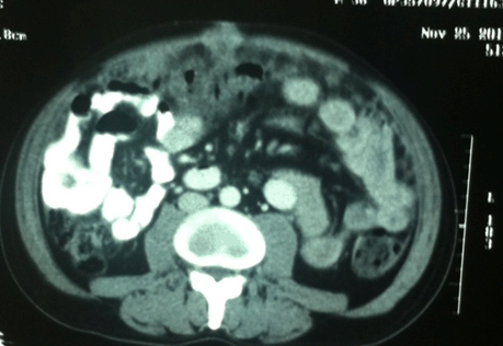
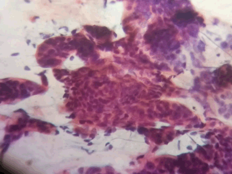
A 39-year-old male presented with a two-month history of swelling over the umbilicus with pain abdomen and anorexia. On examination, the patient was asthenic and dehydrated. His general physical examination revealed mild anemia and pedal edema without any lymphadenopathy. On systemic examination, the abdomen was mildly distended with an approximately 2x2 cm umbilical swelling, without any other visible lump, scar or sinus (Figure 1). On abdominal palpation, a hard, tender umbilical swelling was felt that was arising from the anterior abdominal wall. There was no organomegaly, succession splash or fluid thrill in the abdomen. The rest of the systemic examination did not reveal any abnormality. There was no abnormality on digital per rectal examination. The investigations revealed a low hemoglobin of 7 g/dL and other hematological investigations were within normal limits. Serum CEA level was 15.68 ng/mL. Ultrasound of the abdomen revealed an isoechoic to hyperechoic lesion in anterior abdominal wall of size 4x3.5 cm with minimal ascites. The liver, pancreas, gallbladder and kidneys were normal and there was no para-aortic lymphadenopathy. Computed tomography scan of the abdomen revealed noncircumferential thickening of upper rectum with mesorectal lymphadenopathy. There was a heterogeneous mass lesion in anterior abdominal wall with multiple deposits over the omentum and peritoneum with minimal free fluid (Figure 2). Colonoscopy revealed a proliferative lesion in mid rectum which was confirmed by histopathology as mucinous adenocarcinoma (Figure 3). Fine needle aspiration cytology (FNAC) of the umbilical nodule done showed metastatic deposits of mucinous adenocarcinoma and a diagnosis of Sister Mary Joseph's nodule was made (Figure 4). Patient receiving palliative chemotherapy with regimen FOLFOX-IV and was on regular follow-up till six months. | ||||||
| ||||||
| ||||||
|
| ||||||
| ||||||
|
Discussion
| ||||||
|
The term "Sister Joseph's nodule" was proposed by a British Surgeon, Sir Hamilton Bailey for the umbilical metastasis of an abdominal cancer in 1948. This uncommon pathology has numerous nascent etiologies. Three-fourths malignant umbilical tumors resemble to a "Sister Joseph's nodule" [2]. The incidence of cutaneous metastases from various malignancies reported in a range from 1% to up to 9%, according to autopsy reports. Merely, 10% out of them are presented as an umbilical metastasis [3]. Among all malignant umbilical lesions, 88% are metastatic; the rest are primary skin tumors [2]. Approximate 1–3% of intra-abdominal malignancies, including gastrointestinal and genitourinary may present with umbilical metastasis during the whole course of the disease. Among them, most of the patients 88% metastasized to umbilicus, and the rest to the skin [1] [3]. In patients with a history of known malignancies, as a recurrence either a solitary or synchronous lesion with another systemic dissemination of the disease is a common presentation. The SMJN as an initial presentation of any primary malignancy is reported in up to 30% of the cases, whereas the remainder presents in patients with known history of malignancy [3] [4]. Galvan et al. reported based on a large review of 407 patients over a period of three decades (1966–1997) that 14.6% of umbilical metastatic lesions have colorectal cancers as the primary source of metastasis [5]. Females are predominantly known to have these lesions [2]. Some case reports of SMJN published, as a first presenting feature in rectal cancer [6]. The common primary cancers metastasize to umbilicus are the gastrointestinal (35–65%), and genitourinary (12–35%) tract followed by rarely, hematological malignancies, lung or breast cancers may be in 3–6% of cases. After a thorough investigation of a patient with a metastatic umbilical lesion, 15–30% of patients remain with unknown primary [1] [2]. Most common histology of SMJN is reported adenocarcinoma followed by squamous cell carcinoma, melanoma or sarcoma [7]. The pathogenesis of umbilical metastasis is yet not well understood, various theories, including
The rich arterial supply and venous network flowing cranially and caudally from the umbilicus with lymphatic drainage systems including pelvic and para-aortic lymph nodes favors the deposition of circulating cancer cells to the umbilicus. Traumatic violation of the anterior abdominal wall circulation during invasive procedures like diagnostic laparoscopy or operation for sterilizations especially in women, is also known to be a risk factor for higher dissemination of cancer cells to the anterior abdominal wall [2] [3] [4]. Clinically may present as the first presentation of underlying malignancy or during the progression of known cases of cancer [6] [8] [9]. The differential diagnosis of umbilical nodule includes neoplastic or non-neoplastic lesions such as Paget's disease, angioma, umbilical adenoma (raspberry tumor), umbilical hernia, endometriosis, hypertrophic scar, umbilical granuloma, pilonidal sinus, mycosis psoriasis, and eczema [2] [6]. Tissue diagnosis in the form of fine-needle aspiration cytology is acceptable to establish the diagnosis [10]. Presence of umbilical metastasis usually runs with poor prognosis due to advanced cancer with widespread metastases [2] [3] [6]. However, multimodality therapy including surgery and adjuvant therapy may improve the survival in some patients having good performance status [3]. Rarely, the surgical resection with negative margins with or without reconstruction of abdominal wall defect in isolated umbilical metastatic is a satisfactory curative treatment option, but chemotherapy is usually the mainstay of the treatment [3] [6]. It is dubious to get cured an umbilical metastasis in patients with disseminated cancer. However, systemic chemotherapy like FOLFOX based regimen should be considered as it sometimes gives good response in colorectal cancers. Overall, the prognosis of umbilical metastasis is poor with median survival only about one year [11]. | ||||||
|
Conclusion
| ||||||
|
Sister Mary Joseph nodule is an unusual indicator of occult visceral and other malignancies which can be diagnosed promptly only if physicians are made aware of this rare clinical presentation. It generally signifies disseminated advanced disease with poor prognosis for which aggressive combined modality treatment is required in every individual instance. | ||||||
|
References
| ||||||
| ||||||
|
[HTML Abstract]
[PDF Full Text]
|
|
Author Contributions
Syed Altaf – Substantial contributions to conception and design, Acquisition of data, Analysis and interpretation of data, Drafting the article, Revising it critically for important intellectual content, Final approval of the version to be published Kapil Dev – Analysis and interpretation of data, Revising it critically for important intellectual content, Final approval of the version to be published Jaiprakash Gurawalia – Analysis and interpretation of data, Revising it critically for important intellectual content, Final approval of the version to be published Shiva Kumar – Analysis and interpretation of data, Revising it critically for important intellectual content, Final approval of the version to be published |
|
Guarantor of submission
The corresponding author is the guarantor of submission. |
|
Source of support
None |
|
Conflict of interest
Authors declare no conflict of interest. |
|
Copyright
© 2017 Syed Altaf et al. This article is distributed under the terms of Creative Commons Attribution License which permits unrestricted use, distribution and reproduction in any medium provided the original author(s) and original publisher are properly credited. Please see the copyright policy on the journal website for more information. |
|
|