|

|
 |
|
Case Report
| ||||||
| Role of immunohistochemistry in metastatic clear cell variant of follicular thyroid carcinoma: A case report | ||||||
| Yash Pradeep Vaidya1, Rajan Vaithianathan2, Ramanathan Manickam3, Dhananjay Kotasthane4 | ||||||
|
1MBBS, Postgraduate, General Surgery, Mahatma Gandhi Medical College and Research Institute, Sri Balaji Vidyapeeth University, Pondicherry, India.
2MS (Gen. Surgery), FRCS, Associate Professor, General Surgery, Mahatma Gandhi Medical College and Research Institute, Sri Balaji Vidyapeeth University, Pondicherry, India. 3MS (Gen. Surgery), Professor and Head, General Surgery, Mahatma Gandhi Medical College and Research Institute, Sri Balaji Vidyapeeth University, Pondicherry, India. 4MD (Pathology), Professor and Head, Pathology, Mahatma Gandhi Medical College and Research Institute, Sri Balaji Vidyapeeth University, Pondicherry, India. | ||||||
| ||||||
|
[HTML Abstract]
[PDF Full Text]
[Print This Article]
[Similar article in Pumed] [Similar article in Google Scholar] 
|


| How to cite this article |
| Vaidya YP, Vaithianathan R, Manickam R, Kotasthane D. Role of immunohistochemistry in metastatic clear cell variant of follicular thyroid carcinoma: A case report. Int J Case Rep Images 2017;8(3):196–200. |
|
Abstract
|
|
Introduction:
Clear cell variant of follicular thyroid carcinoma with synchronous bony metastasis and a normal thyroid stimulating hormone level, is an extremely rare condition.
Case Report: A 55-year-old male was presented to us with a painful swelling in the right arm. The biopsy showed clear cell adenocarcinoma, raising concerns for a metastatic renal cell carcinoma. Computed tomography scan of abdomen failed to show any renal lesions. A detailed physical examination revealed a small nodule of the right thyroid lobe. Fine needle aspiration cytology (FNAC) of the nodule was reported as follicular neoplasm. A right hemithyroidectomy and the subsequent completion thyroidectomy showed clear cell type of follicular thyroid carcinoma. Immunohistochemistry for thyroglobulin (Tg) further confirmed the diagnosis. Conclusion: Clear cell variant of follicular thyroid carcinoma is a very rare condition, hence a high index of suspicion is essential for diagnosis. The importance of performing a detailed physical examination cannot be more emphasized as small thyroid lesion like in this case can be easily missed, leading to a delay in diagnosis. | |
|
Keywords:
Clear cell variant, Follicular thyroid carcinoma, Thyroglobulin, Thyroidectomy
| |
|
Introduction
| ||||||
|
Follicular thyroid carcinoma is the second most common type of thyroid cancer, comprising around 10–15% of thyroid malignancies. It has been histologically classified into oncocytic and clear cell types by the World Health Organization [1]. Follicular thyroid carcinoma with clear cell change is rare, and only some cases have been reported in literature [2]. This can be a diagnostic problem as metastatic clear cell carcinoma to the bones is usually attributed to a renal primary. Moreover, the most common cause for metastatic lesion in the thyroid is also renal cell carcinoma [3]. This clinical scenario may prove very challenging in arriving at an accurate diagnosis and for optimum management. | ||||||
|
Case Report
| ||||||
|
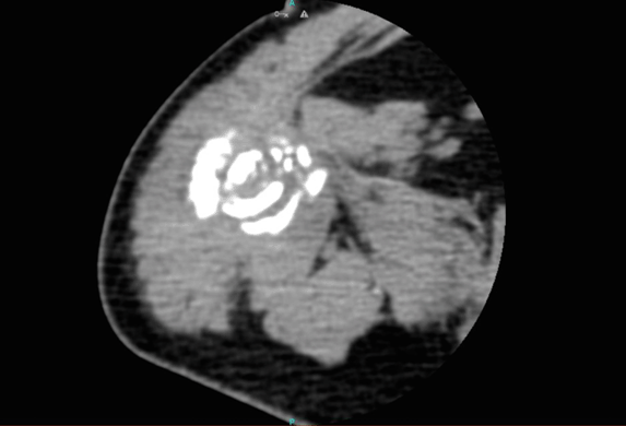
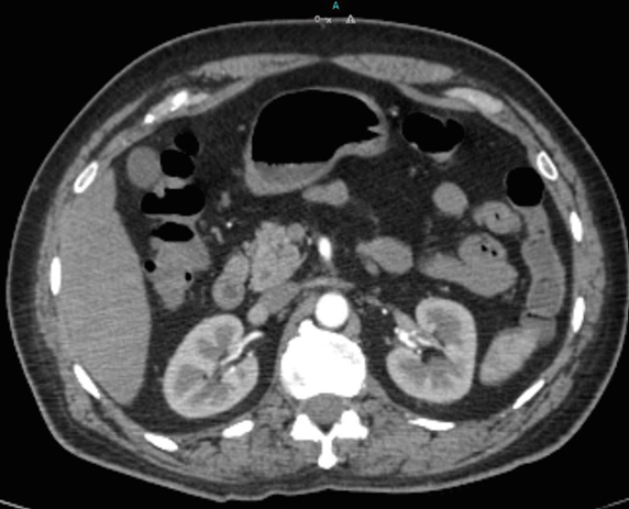
A 55-year-old male initially presented with pain and difficulty in moving his right arm after a fall. No significant past medical or family history was noted. On examination, he had a hard, 6x4 cm swelling over the upper third of the humerus. X-ray of the right humerus showed a soft tissue mass and a lytic lesion causing pathological fracture with varus angulation. Magnetic resonance imaging scan showed an intramedullary lesion in the bone with soft tissue extension, suggestive of a primary bone malignancy (Figure 1). An open biopsy of the lesion revealed clear cell adenocarcinoma, probably of renal origin (Figure 2). However, immunohistochemistry staining using vimentin, CD 10, cytokeratin and S100 were equivocal. Computed tomography scan of abdomen was normal (Figure 3). On further examination, a small nodule was palpable in the right lobe of the thyroid. Thyroid function tests were within normal limits. Ultrasound of the neck demonstrated a 2x1 cm, hypoechoic nodule in the right lobe. Ultrasound guided FNAC was suggestive of a follicular neoplasm. A U-slab was applied for the fracture in right humerus. The patient underwent an initial right hemithyroidectomy. Preoperatively, the patient was explained in detail about the need for this procedure as well as for completion thyroidectomy if malignancy was confirmed. Histopathology proved it to be clear cell variant of follicular thyroid carcinoma, with tumor cells invading into vascular spaces, features very similar to the biopsy from the right humerus (Figure 4). He subsequently underwent completion thyroidectomy to remove the left lobe and this also showed similar features. Immunohistochemistry was strongly positive for thyroglobulin and negative for prior mentioned markers, thus confirming the lesion to be a thyroid primary rather than a metastatic lesion from the kidneys. The patient made an uneventful recovery, with no symptoms of hypocalcaemia. He was later referred to the regional Radiation Oncology Centre for radioactive iodine (RAI) treatment and planned for a RAI scan (both thyroid and whole body). We plan to monitor the patient regularly with serum thyroglobulin levels. | ||||||
| ||||||
|
| ||||||
| ||||||
| ||||||
|
Discussion
| ||||||
|
Most follicular thyroid carcinomas are asymptomatic and diagnosed late. Thyroid scintigraphy usually shows a cold nodule. They typically present as solitary lesions, and very rarely involve both the lobes. Distant metastasis is common when there is capsular or vascular invasion [1]. Follicular thyroid carcinoma commonly spreads by the hematogenous route. Around 10–15% of patients have distant metastasis, occurring more often in tumors larger than 2 cm size [4]. The most common sites of distant metastases are the bones (with typical lytic lesions) and lungs followed by the brain, liver, bladder and skin [5]. The incidence of metastatic disease is higher in the oncocytic variant than the clear cell type [6]. The clear cell appearance has been attributed to either accumulation of vesicles or glycogenation, thyroglobulin accumulation and hypertrophy of the Golgi complex due to excessive thyroid stimulating hormone stimulation [1] [7] [8]. Presence of clear cell features in follicular thyroid carcinoma very rare in patients with a normal thyroid stimulating hormone value as noted in this patient. Almost 25–30% patients with renal cell carcinoma have distant metastasis at the time of diagnosis [9]. Hence this is usually the first differential diagnosis in such cases. When metastatic lesions in the bone and the thyroid exhibit clear cell changes, it becomes difficult to differentiate metastatic renal cell carcinoma from a primary clear cell follicular thyroid carcinoma [10]. In such cases, immunohistochemistry plays an important role in clinching the origin of the tumor cells, as we found in our case. Clear cell renal cell carcinoma usually stains positive for vimentin, cytokeratin antibodies (AE1/AE3), CD10, renal cell carcinoma marker (RCCM), PAX2, PAX8, and carbonic anhydrase IX (CAIX), and stains negative for cytokeratins (CK7, CK 20), high-molecular-weight cytokeratin (HMWCK), CD 117 and parvalbumin [11]. | ||||||
|
Conclusion
| ||||||
|
Clear cell variant of follicular thyroid carcinoma is a very rare condition, hence a high index of suspicion is essential for diagnosis. Metastatic carcinomas exhibiting clear cell changes are commonly mistaken for a spread from a renal cell carcinoma . The importance of performing a detailed physical examination cannot be more emphasized as small thyroid lesion like in this case can be easily missed, leading to a delay in diagnosis. Immunohistochemistry is the definitive way of differentiating clear cell carcinomas arising from thyroid and the kidneys. | ||||||
|
Acknowledgements
| ||||||
|
We would like to thank Prof. N. Ananthakrishnan, Dean of PG studies and Research, for his valuable contribution during the discussion of this case and treatment planning. | ||||||
|
References
| ||||||
| ||||||


|
[HTML Abstract]
[PDF Full Text]
|
|
Author Contributions
Yash Pradeep Vaidya – Substantial contributions to conception and design, Acquisition of data, Analysis and interpretation of data, Drafting the article, Revising it critically for important intellectual content, Final approval of the version to be published Rajan Vaithianathan – Substantial contributions to conception and design, Drafting the article, Revising it critically for important intellectual content, Final approval of the version to be published Ramanathan Manickam – Substantial contributions to conception and design, Drafting the article, Revising it critically for important intellectual content, Final approval of the version to be published Dhananjay Kotasthane – Substantial contributions to conception and design, Drafting the article, Revising it critically for important intellectual content, Final approval of the version to be published |
|
Guarantor of submission
The corresponding author is the guarantor of submission. |
|
Source of support
None |
|
Conflict of interest
Authors declare no conflict of interest. |
|
Copyright
© 2017 Yash Pradeep Vaidya et al. This article is distributed under the terms of Creative Commons Attribution License which permits unrestricted use, distribution and reproduction in any medium provided the original author(s) and original publisher are properly credited. Please see the copyright policy on the journal website for more information. |
|
|
|
About The Authors
| |||
| |||
| |||
| |||
| |||