| |
|
|
|
Case Report
| ||||||
| Perforated gangrenous cholecystitis with concurrent Clostridium perfringens bacteraemia masquerading as adenomyomatosis of the gallbladder: A case report | ||||||
| Natalie LY Ngu1, Alexander Olaussen1,2,7, Jessica Wong3, Hayden Snow3, Mark Cullinan4,6, Paul J. Sitzler4,5 | ||||||
|
1Intern, General Surgery, Sandringham District and Memorial Hospital, Alfred Health, Melbourne, Victoria, Australia.
2Adjunct Senior Lecturer Monash University, Department of Community Emergency Health and Paramedic Practice. 3Registrar, General Surgery, Sandringham District and Memorial Hospital, Alfred Health, Melbourne, Victoria, Australia. 4Consultant Surgeon, General Surgery, Sandringham District and Memorial Hospital, Melbourne, Victoria, Australia. 5Head of Unit, General Surgery, Sandringham District and Memorial Hospital, Melbourne, Victoria, Australia. 6Senior Lecturer, Monash University, Department of Surgery, School of Clinical Sciences. 7National Trauma Research Institute, The Alfred Hospital, Melbourne, Australia. | ||||||
| ||||||
|
[HTML Abstract]
[PDF Full Text]
[Print This Article]
[Similar article in Pumed] [Similar article in Google Scholar] 
|
| How to cite this article |
| Natalie LY Ngu, Olaussen A, Wong J, Snow H, Cullinan M, Sitzler PJ. Perforated gangrenous cholecystitis with concurrent Clostridium perfringens bacteraemia masquerading as adenomyomatosis of the gallbladder: A case report. Int J Case Rep Images 2017;8(3):179–183. |
|
Abstract
|
|
Introduction:
Clostridium perfringens (C. perfringens) is an unusual cause of bacteraemia in the healthy, immunocompetent host. Similarly, acalculous cholecystitis is rare in the absence of critical illness or preceding trauma. We present, to the best of our knowledge, the first documented case of concurrent C. perfringens bacteraemia and acalculous cholecystitis in a previously well human.
Case Report: An apparently healthy 56-year-old male was presented with sepsis of unknown origin, and was treated for a respiratory infection and incidentally found to have gallbladder mural thickening on a computed tomography (CT) chest scan. An abdominal ultrasound (USG) demonstrated adenomyomatosis of the gallbladder, without evidence of acute cholecystitis or gallstones. The initial blood sample hemolyzed, however, subsequent specimens showed raised inflammatory markers, neutrophilia and thrombocytopenia. The patient continued to deteriorate clinically and biochemically. At 30 hours from presentation, blood cultures demonstrated a C. perfringens bacteraemia and intravenous antibiotics were commenced. Following these findings and the development of right upper quadrant abdominal pain, biliary sepsis was suspected and the patient taken to theatre. During laparoscopic cholecystectomy, a perforated and gangrenous gallbladder was identified and the intraoperative cholangiogram demonstrated no retained stones. Acute gangrenous cholecystitis was confirmed on histopathology. Postperatively, the patient recovered quickly and was discharged with oral antibiotics. Conclusion: We present a case of acalculous cholecystitis in a patient with an unusual clinical presentation and lack of positive imaging findings. In this setting, the need for definitive surgical intervention and clinical suspicion of cholecystitis was recognized with the finding of C. perfringens bacteraemia despite imaging suggesting adenomyomatosis. This case highlights that acalculous cholecystitis can occur in patients without risk factors, and can be complicated by atypical bacteraemia, even in previously healthy individuals. | |
|
Keywords:
Acalculous cholecystitis, adenomyomatosis, Bacterae-mia, Clostridium perfringens
| |
|
Introduction
| ||||||
|
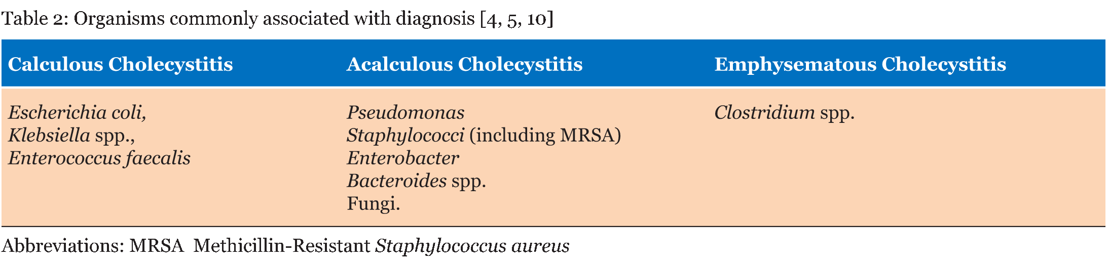
C. perfringens is a potentially dangerous gram positive anaerobic rod, causing disease through toxin release. The incidence has been estimated to be less than 2 per 100,000 [1] and mortality rates have been reported between 27% and 44% [1]. Recognized sources of C. perfringens bacteraemia include wound contamination and iatrogenic bowel leakage, and can be complicated by hemolysis and overwhelming infection if treatment is delayed [2]. Acute cholecystitis commonly presents with a syndrome of right upper quadrant abdominal pain, fever and leukocytosis, and is usually attributed to gallstones [3]. Common organisms associated with cholecystitis include Escherichia coli, Klebsiella spp. and Enterococcus faecalis [4]. Acalculous cholecystitis has a similar clinical picture but in the absence of gallstones. It accounts for 2–15% [5] of acute cholecystitis cases and often presents in critically ill patients or with severe systemic stress e.g., trauma, major surgery, shock or burns [6]. Acalculous cholecystitis is more frequently associated with Pseudomonas, Staphylococci including methicillin-resistant Staphylococcus aureus, Enterobacter, Bacteroides spp. and fungi [4]. We present a diagnostic dilemma of concurrent acalculous cholecystitis inaccurately identified on ultrasound and C. perfringens bacteraemia in a previously healthy individual, with implications for future diagnosis. | ||||||
|
Case Report
| ||||||
|
A 56-year-old male presented to the emergency department with a history of one day of severe chest and epigastric pain, fevers and rigors. His medical history included ischemic heart disease, peptic ulcer disease and gastroesophageal reflux disease. On examination, he was febrile (39.4°C), demonstrated tachycardia of 110 beats per minute, tachypnea of 26 breaths per minute, an oxygen saturation of 92% on one litre of supplemental oxygen and was normotensive. His chest and abdominal examinations were normal. An initial blood sample was hemolyzed prior to analysis. Subsequent blood tests revealed a mild neutrophilia, raised C-reactive protein (CRP) and thrombocytopenia (Table 1). All other blood levels including liver function tests were normal. Since a chest X-ray demonstrated no abnormality, a CT pulmonary angiogram was performed for suspected pulmonary embolism, however, the only salient findings were gallbladder wall thickening and a small volume of pericholecystic fluid. An abdominal USG suggested adenomyomatsis of the gallbladder without features of cholecystitis. Gallstones were not visualized on either scan. Empirical intravenous antibiotics (ceftriaxone 1 g and azithromycin 500 mg) were commenced. The patient continued to be febrile overnight with mild right upper quadrant abdominal pain developing. The following morning, the CRP had increased and the platelet count had fallen further (Table 1). Preliminary blood cultures taken at time of presentation, demonstrated a C. perfringens bacteraemia at 30 hours from presentation, and antibiotics were changed to intravenous tazobactam/piperacillin 4.5 g and clindamycin 600 mg. This positive blood culture result and the location of abdominal pain greatly increased suspicion for biliary sepsis, and the patient was taken to theatre. Intraoperatively, a perforated and gangrenous gallbladder was removed laparoscopically and a cholangiogram demonstrated no filling defects. The patient received a single pack of pooled, irradiated platelets, due to refractory bleeding from the gallbladder fossa. Postoperatively, the thrombocytopenia had improved and the patient was discharged with oral antibiotics (amoxicillin/clavulanate 875/125 mg). Acute gangrenous cholecystitis without gallstones was confirmed on histopathology (Figure 1). At a two-week follow-up appointment, all blood abnormalities had resolved to within normal range (Table 1). | ||||||
| ||||||
| ||||||
| ||||||
|
Discussion
| ||||||
|
This case represents a clinical dilemma given the unusual presentation and difficult pathway to diagnosis. The diagnosis of acalculous cholecystitis was finally made based on intraoperative findings of a perforated and gangrenous gallbladder, the normal cholangiogram and the subsequent histopathologic analysis of the specimen. As outlined in Table 2, the commonly associated organisms vary between calculous, acalculous and emphysematous cholecystitis. Although Clostridium spp. has been associated with secondary emphysematous acalculous cholecystitis in critically ill patients or following antibiotics [3], no features suggestive of emphysematous acalculous cholecystitis including air in the gallbladder lumen or wall were seen on imaging in this case. In addition, the absence of a preceding physiological stress or immunocompromise is unusual in the development of both acalculous cholecystitis and C. perfringens bacteraemia. Although no other case of acalculous cholecystitis and C. perfringens bacteraemia has been reported in human literature, there is a similar published case of a pig with undifferentiated sepsis, a necrotic gall bladder without gallstones on autopsy and hemolysis of initial blood samples [6]. A human case of C. perfringens bacteraemia with clinical suspicion of a biliary source, was attributed to gallstones, which were confirmed on endoscopic retrograde cholangiopancreatography [7]. These examples highlight the atypical presentation in our patient and the need to consider all clinical features when faced with a similar diagnostic dilemma. The significance of the patient's thrombocytopenia remains unclear, however, a corresponding phenomenon has been reported in a retrospective cohort study of 93 patients with C. perfringens [1]. Additionally, hemolysis of an initial blood sample has been reported in another case of C. perfringens bacteraemia [8]. This may be attributed to toxin release [9]. However further exploration of these two associations is needed. | ||||||
|
Conclusion
| ||||||
|
C. perfringens bacteraemia is a rare and potentially serious condition. Acalculous cholecystitis can occur despite abdominal ultrasonography suggesting an alternative diagnosis. This case highlights that acalculous cholecystitis can occur in patients without risk factors, and can be complicated by atypical bacteraemia, even in previously healthy individuals. | ||||||
|
Acknowledgements
| ||||||
|
Dr Rhoda Cameron, Consultant Pathologist, Anatomical Pathology, Alfred Health, Melbourne, Victoria. Informed, written consent for the synthesis and publication of this case report was obtained from the patient, with thanks. | ||||||
|
References
| ||||||
| ||||||
|
[HTML Abstract]
[PDF Full Text]
|
|
Author Contributions
Natalie LY Ngu – Substantial contributions to conception and design, Acquisition of data, Analysis and interpretation of data, Drafting the article, Revising it critically for important intellectual content, Final approval of the version to be published Alexander Olaussen – Substantial contributions to conception and design, Acquisition of data, Analysis and interpretation of data, Drafting the article, Revising it critically for important intellectual content, Final approval of the version to be published Jessica Wong – Substantial contributions to conception and design, Drafting the article, Revising it critically for important intellectual content, Final approval of the version to be published Hayden Snow – Substantial contributions to conception and design, Revising it critically for important intellectual content, Final approval of the version to be published Mark Cullinan – Substantial contributions to conception and design, Revising it critically for important intellectual content, Final approval of the version to be published Paul Sitzler – Substantial contributions to conception and design, Revising it critically for important intellectual content, Final approval of the version to be published |
|
Guarantor of submission
The corresponding author is the guarantor of submission. |
|
Source of support
None |
|
Conflict of interest
Authors declare no conflict of interest. |
|
Copyright
© 2017 Natalie LY Ngu et al. This article is distributed under the terms of Creative Commons Attribution License which permits unrestricted use, distribution and reproduction in any medium provided the original author(s) and original publisher are properly credited. Please see the copyright policy on the journal website for more information. |
|
|