| |
|
|
|
Case Report
| ||||||
| A successfully-treated case of penetrating facial trauma | ||||||
| Tetsuya Yumoto1, Atsuyoshi Iida1, Kohei Tsukahara1, Hiromichi Naito1, Michihisa Terado1, Keiji Sato1, Isao Date2, Atsunori Nakao1 | ||||||
|
1Department of Emergency and Critical Care Medicine, Okayama University Graduate School of Medicine, Dentistry, and Pharmaceutical Sciences.
2Department of Neurological Surgery, Okayama University Graduate School of Medicine, Dentistry, and Pharmaceutical Science. | ||||||
| ||||||
|
[HTML Abstract]
[PDF Full Text]
[Print This Article]
[Similar article in Pumed] [Similar article in Google Scholar] 
|
| How to cite this article |
| Yumoto T, Iida A, Tsukahara K, Naito H, Terado M, Sato K, Date I, Nakao A. A successfully-treated case of penetrating facial trauma. Int J Case Rep Images 2017;8(2):120–123. |
|
Abstract
|
|
Introduction:
Penetrating intracranial injury caused by a metal bar is rare and often causes severe damage without fast treatment.
Case Report: A 59-year-old male fell from a height of four feet onto upward-pointed metal bars and was transferred to our emergency department after suffering an accidental penetration of a metal bar through his face. Computed tomography (CT) scans were conducted; however, the image resolution was unsatisfactory and the amount of brain damage could not be evaluated due to serious artifacts associated with the metal bar. After removing the foreign body, follow-up CT scn revealed increased hemorrhage in the frontal lobe. Emergency craniotomy and removal of the hematoma followed. Conclusion: Artifacts from a penetrating metal bar on CT scan often hide actual brain damage along the trajectory of penetration. Emergent surgical intervention and early follow-up CT scan is necessary for any chance to save the patient's life. In these cases, proper examination, radiological tests, and early decision-making using a multidisciplinary strategy provide optimal outcomes. | |
|
Keywords:
Artifacts, Intracranial stab injury, Metal bar, Transorbital brain injury
| |
|
Introduction
| ||||||
|
Penetrating intracranial stab injuries caused by metal bars are very rare among ordinary people and often cause severe damage without swift treatment [1]. Penetrating trauma to the face presents a broad spectrum of injuries and treatment challenges due to potential injuries that could occur when the penetrating object is removed. Immediate complications of transfacial penetrating trauma include cerebral contusion, intracerebral hematoma, pneumocephalus, intraventricular hemorrhage, cranial nerve damage, severe permanent neurological damage, and brain stem and cerebrovascular injury [2]. Delayed complications include cerebrospinal fluid fistula, pneumocephalus, orbital cellulitis, carotid-cavernous sinus fistula, central nervous system infections, traumatic aneurysm, and delayed intracranial hemorrhage [3]. Computed tomography (CT) scan is usually the first-line radiologic assessment in the emergency room for patients with head injuries. However, artifacts caused by the penetrating object on CT scan often hide the extent of actual brain damage along the trajectory of penetration and treatment of the brain insult may be delayed. Infections can easily complicate penetrating craniocerebral injuries, subdural empyemas, or brain abscesses. Treatment strategies for facial penetrating injury, including prevention and proper management of infectious complications, should be decided with input from multidisciplinary experts such as neurosurgeons, otolaryngologists, and ophthalmologists. This case report may provide guidelines for effective treatment of an intracranial penetrating injury, which may help emergency physicians. | ||||||
|
Case Report
| ||||||
|
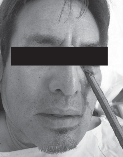
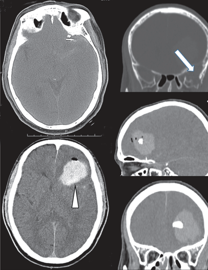
A 59-year-old male with a penetrating injury to his face was presented to our department. The patient had fallen from a height of approximately four feet and impaled himself on a metal bar (1 cm in diameter). The iron bar, which had been cut, was still embedded in the right side of his face, approximately 3 cm below his left eye, penetrating his maxillofacial region (Figure 1). The patient did not experience any loss of consciousness and paramedics reported no significant blood loss at the scene. On arrival, his Glasgow Coma Scale score was E4V5M6, the airway was clear, and cervical spine injury was excluded. His vital signs included a heart rate 78 beats/min, blood pressure 142/84 mmHg, respiratory rate 18 breaths/min, and axillary temperature 36.3°C. The pupils were equal in size, round, and reactive to light. His vision was normal bilaterally. No motor or sensory deficits were present on the neurological examination. Computed tomography scan of the brain showed marked artifacts as bright and dark streaks over the trajectory of the metal bar without obvious brain contusion hemorrhage. Therefore, CT scan was again performed after the foreign metallic body was carefully removed with X-ray guided. Right mandibular and sphenoidal bone fracture and intraparenchymal hematoma in the frontal lobe with pneumocephalus caused by direct compaction of the metal bar were noted (Figure 2). Following imaging, the patient became drowsy and was emergently transported to the operating room. Under general anesthesia, emergent craniotomy and hematoma removal was performed 90 minutes after removal of the bar. Broad-spectrum antibiotic treatment with ceftriaxone was started. The patient recovered well from the surgery and the wounds healed without incident or cerebrospinal fluid leak. Ophthalmic examination revealed normal vision 10 days postoperatively. No facial paralysis was noted. | ||||||
| ||||||
| ||||||
|
Discussion
| ||||||
|
Penetrating craniofacial injury is rare, accounting for 0.4% of all head injuries [4]. Most penetrating brain injuries in civilian emergency practice are missile injuries. Most non-missile penetrating brain injuries are caused by knives, pens, and chopsticks. The face has protective reflexes that help divert it from oncoming objects. The face has a smaller surface area than the trunk or extremities. Furthermore, the structure of the face and cranium are suited to absorb shocks, owing to the presence of resistant pillars, buttresses, and the presence of pneumatized cavities. Therefore, in general, penetrating facial injuries result in less morbidity to the patient. A penetrating facial injury may not seem serious at first glance, but there may be deeper intracranial injury or injury to the orbit or dura that can be overlooked. The concentrated force of the small area at the tip of the metal bar may enable penetration into the cranial bone. The mechanism of vascular and neuronal injuries caused by cranial stab wounds may differ from that caused by other types of head trauma. Unlike high-energy accidents, no diffuse shearing injury to the brain occurs. Therefore, a patient with intracranial stab wounds usually has a better prognosis than a patient with penetrating injuries caused by an object with a higher kinetic energy [5]. Radiology of the cranium is helpful in evaluating the intracranial course of metallic materials, but the radiodensity of wood is almost the same as that of the brain and soft tissue, so it is difficult to detect. Although it may be limited for evaluating plastic or wood, head CT scan is the most valuable test for first line evaluation of foreign objects and assessing the extent of injury. However, in many cases, optimal evaluation cannot be performed on cerebral CT scan due to the presence of artifacts caused by the object. Angiography is advocated by some authors for possible cerebral vascular injuries in patients after penetrating head injuries to rule out unexpected vascular lesions. Arteriovenous fistulae and traumatic aneurysms are the most important findings, since they are seldom clinically evident. Delaying carotid angiography until the start of the second week has previously been proposed to allow for better visualization of these complications. However, traumatic aneurysms can burst at any time post injury, and the mortality from a second hemorrhage is unacceptably high [6] [7]. Management of patients with penetrating brain injuries should follow fundamental surgical principles, including removing the object under direct vision in order to reduce further brain tissue damage by the foreign body catching on the bone fragments. The most appropriate management in the pre-hospital environment is to leave the penetrating object in situ and move the patient to the trauma center. Key strategies for emergency physicians and medical technicians are to stabilize the object to prevent movement and conduct serial neurological examinations. Having a systematic method to evaluate and manage such injuries is important. Following removal of the foreign body, thorough debridement with removal of all involved skull bone and retained fragments, evacuation of the hematoma followed by careful hemostasis along the trajectory, and meticulous dural closure to reduce the chance of cerebrospinal fluid fistula are mandatory. Prevention of infection needs to be addressed. Patients should be given tetanus shots and preoperative and postoperative antibiotics, if indicated. Some reports suggest that although penetrating craniofacial injuries with a wooden bar, which can affect extensively by broken fragments, usually require surgical intervention, injuries with a metal bar can be managed without surgical intervention with good outcomes [8]. It is imperative to leave the foreign body in place until adequate preoperative planning has been completed and subsequent removal should only be attempted under direct visualization in an operating room. Premature removal without adequate preparation can put the patient at risk for further injury or fatal bleeding if the object has caused a tamponade of the bleeding [9]. Our patient presented with corresponding brain hemorrhages with a great mass obscured on the follow-up CT scan, indicating that bleeding occurred after removal of the object. Based on this experience, we recommend immediate surgical decompression with early follow-up CT to ascertain the extent of brain damage and monitor patients for possible delayed events. Prognosis depends on the affected brain area and the involvement of major vessel injuries. Complications and sequelae after penetrating brain injury are including life-threatening hemorrhage, local trauma to the brain and its vasculature, brain abscesses, meningitis, cerebrospinal fluid leakage, and neurological deficits. Epilepsy, behavioral changes, and psychological issues could be attributed to frontal lobe injury [10]. In our patient, proper assessment of the injury and management decisions were made with input from a multidisciplinary team of clinicians in neurosurgery, ophthalmology, otolaryngology and plastic surgery. | ||||||
|
Conclusion
| ||||||
|
Transorbital brain injury caused by a metal bar is an uncommon but potentially fatal event unless managed appropriately. Computed tomography scan of the brain showed marked artifacts over the trajectory of the metal bar. Emergent surgical intervention and early follow-up computed tomography scans are necessary to have any chance to save the patient's life. | ||||||
|
References
| ||||||
| ||||||
|
[HTML Abstract]
[PDF Full Text]
|
|
Author Contributions
Tetsuya Yumoto – Substantial contributions to conception and design, Acquisition of data, Analysis and interpretation of data, Drafting the article, Revising it critically for important intellectual content, Final approval of the version to be published Atsuyoshi Iida – Analysis and interpretation of data, Revising it critically for important intellectual content, Final approval of the version to be published Kohei Tsukahara – Analysis and interpretation of data, Revising it critically for important intellectual content, Final approval of the version to be published Hiromichi Naito – Analysis and interpretation of data, Revising it critically for important intellectual content, Final approval of the version to be published Michihisa Terado – Analysis and interpretation of data, Revising it critically for important intellectual content, Final approval of the version to be published Keiji Sato – Analysis and interpretation of data, Revising it critically for important intellectual content, Final approval of the version to be published Isao Date – Substantial contributions to conception and design, Analysis and interpretation of data, Revising it critically for important intellectual content, Final approval of the version to be published Atsunori Nakao – Substantial contributions to conception and design, Acquisition of data, or analysis and interpretation of data, Drafting the article or revising it critically for important intellectual content, Final approval of the version to be published |
|
Guarantor of submission
The corresponding author is the guarantor of submission. |
|
Source of support
None |
|
Conflict of interest
Authors declare no conflict of interest. |
|
Copyright
© 2017 Tetsuya Yumoto et al. This article is distributed under the terms of Creative Commons Attribution License which permits unrestricted use, distribution and reproduction in any medium provided the original author(s) and original publisher are properly credited. Please see the copyright policy on the journal website for more information. |
|
|