| |
|
|
|
Case Report
| ||||||
| Liver disease masquerading as primary cardiopulmonary disease: Hepatopulmonary syndrome as a result of idiopathic cirrhosis | ||||||
| Bartholameuz Nuwan Aravinda1, Ratnatilaka Asoka2, Sadikeen Aflah3 | ||||||
|
1MBBS, Registrar-Medicine, National Hospital, Colombo, Sri Lanka.
2MBBS, MD, Consultant Physician, National Hospital, Colombo, Sri Lanka. 3MBBS, MD, Consultant Chest Physician, Central Chest Clinic, Colombo, Sri Lanka. | ||||||
| ||||||
|
[HTML Abstract]
[PDF Full Text]
[Print This Article]
[Similar article in Pumed] [Similar article in Google Scholar] 
|
| How to cite this article |
| Bartholameuz NA, Ratnatilaka A, Sadikeen A. Liver disease masquerading as primary cardiopulmonary disease: Hepatopulmonary syndrome as a result of idiopathic cirrhosis. Int J Case Rep Images 2017;8(2):116–119. |
|
Abstract
|
|
Introduction:
Liver disease and portal hypertension can be associated with pulmonary vascular complications including hepatopulmonary syndrome and portopulmonary hypertension. Hepatopulmonary syndrome is characterized by arterial hypoxemia and pulmonary vascular dilatations. Exertional dyspnea, platypnea-orthodeoxia, cyanosis and digital clubbing are commonly found symptoms and signs in hepatopulmonary syndrome.
Case Report: Here we discuss a patient with chronic liver disease whose initial presentation was hepatopulmonary syndrome with progressive exertional dyspnea, cyanosis and clubbing. Diagnosis of hepatopulmonary syndrome was made through a constellation of findings in blood gas analysis, contrast echocardiography with biochemical and ultrasound evidence of liver disease applied to standard criteria; having excluded other possible causes. Conclusion: Hepatopulmonary syndrome should be suspected in any patient with liver disease and hypoxia. It should also be formulated in the differential diagnosis of a patient with otherwise unexplained exertional dyspnea and cyanosis with digital clubbing. | |
|
Keywords:
Cirrhosis, Cyanosis, Digital clubbing, Hepatopulmonary syndrome
| |
|
Introduction
| ||||||
|
Cyanosis indicates the presence of deoxygenated haemoglobin of ≥5 g/dL in arterial blood. Causes of cyanosis include intracardiac right-left shunts, lung diseases and pulmonary arteriovenous malformation (AVM). Clubbing is due to fibrovascular proliferation in nail beds, mediated by platelet derived growth factor released from megakaryocytes or platelet emboli which do not reach nail bed unless there is pulmonary capillary damage or intracardiac shunts [1]. Here we discuss a rare cause of dyspnea, cyanosis and clubbing associated with cirrhosis which mimicked primary cardiopulmonary disease. | ||||||
|
Case Report
| ||||||
|
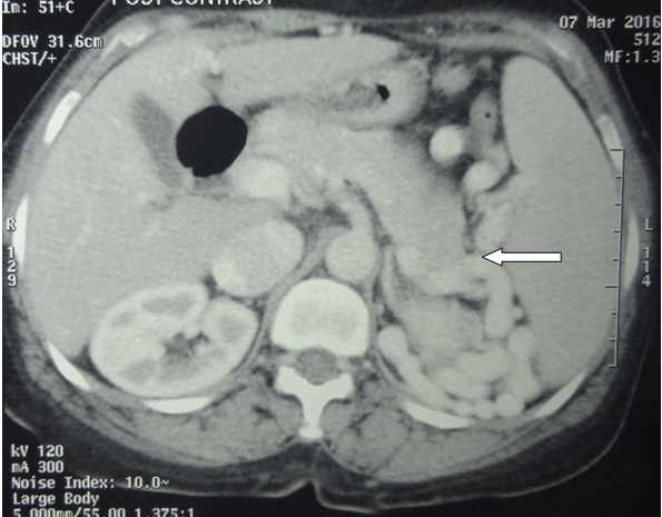
A 56-year-old female was admitted with a history of progressive shortness of breath on exertion for 6 months duration. She had severe central cyanosis with gross clubbing. Mild icterus, palmar erythema and prominent peripheral pulses were also noted. Respiratory rate was 24/min. Systemic examination was normal. Arterial oxygen saturation (SpO2) lying supine was 80% and on standing was 76%. Investigations revealed leucopenia (4.31x10³/µL), thrombocytopenia (31x103/µL), and hemoglobin 15.6 g/dL with macrocytosis in peripheral blood smear. Arterial blood gas (ABG) analysis in supine position showed pH 7.43, PCO2 28.6 mmHg, PaO2 56 mmHg, HCO3- 19.4 mmol/L and Alveolar-arterial oxygen gradient of 58 mmHg. PaO2 dropped to 50.2 mmHg upon standing. Total serum protein was normal (67 g/dL) with reversed serum albumin: globulin ratio (0.6). Serum bilirubin was elevated (42.8 µmol/L). PT was 17.4 s (INR 1.26) and APTT was 46 s (26 s–40 s). Liver enzymes were normal (AST 40 U/L, ALT 22 U/L, and ALP 107 U/L). Abdominal ultrasound detected coarse echo texture in a normal size liver, splenomegaly (12.5 cm; normal <12 cm), portal vein diameter of 1.4 cm (<1.3 cm) without ascites or pleural effusion. Esophagogastroduodenoscopy showed mild portal hypertensive gastropathy and no varices. Hepatitis B and C serology were negative. Serum ferritin was 172 ng/mL. Serum ceruloplasmin levels were normal and there were no Kayser-Fleischer rings. ANA and anti-mitochondrial antibodies were negative. Lipid profile was normal. TSH, Free T4, and HbA1c were normal. High resolution computed tomography of the chest (HRCT) revealed normal lung parenchyma (Figure 1). Contrast-enhanced computed tomography (CECT) of the chest and pulmonary angiogram detected pulmonary venous congestion and dilated venous collaterals without evidence of pulmonary hypertension or AVM. Gross splenorenal collaterals were noted (Figure 2). No evidence of portal vein thrombosis was found. Transthoracic echocardiogram was normal. Transesophageal echocardiography detected a small patent foramen ovale (PFO) but bubble contrast study excluded functional PFO. There was indirect evidence of pulmonary capillary dilatation by detecting micro-bubbles in left atrium after three cardiac cycles following the appearance of bubbles in the right atrium. Her lung function tests were normal (FEV1 94.1%; FVC 99.5%; FEV1: FVC 94.6%, VC 88.1%) except for low DLCO (44%) reflecting a diffusion-perfusion defect due to pulmonary vascular dilatation and hyperdynamic circulation. This is a case with hypoxia, clubbing, cirrhosis and portal hypertension with evidence of intra-pulmonary vascular dilatation (IPVD) confirming the diagnosis of hepatopulmonary syndrome (HPS). | ||||||
| ||||||
| ||||||
|
Discussion
| ||||||
|
In this case, cyanosis was due to dilatation of pulmonary vessels leading to ventilation perfusion mismatch causing hypoxia. Aetiology of pulmonary vascular dilatation in cirrhosis is thought to relate to an increase in pulmonary NO by means of both endothelial and inducible NO synthase (eNOS and iNOS) [2] [3][4]. An increased hepatic production of vasoconstrictor Endothelin-1 (ET-1) stimulates the production of ETB receptors in pulmonary microcirculation. ETA causes vasoconstriction while ETB causes vasodilatation through increase in eNOS activity [2] [3] [4]. Portal hypertension weakens intestinal mucosal barrier due to impaired drainage. It allows increased enteral translocation of bacteria and endotoxins which stimulate release of vasoactive substances like TNF-a. This leads to increased pulmonary sequestration of macrophages and local production of pro-inflammatory mediators causing an increase in iNOS activity and NO production [2] [3] [4]. Normal pulmonary functions and HRCT of the chest excluded chronic parenchymal lung disease. Transesophageal echocardiography with bubble contrast study could not reveal any functional intra-cardiac shunts but indicated presence of pulmonary vascular dilatation in the absence of AVM. These micro-bubbles are larger than normal pulmonary capillary diameter (8–15 µm) and cannot pass through normal pulmonary capillaries [2]. Evidence of pulmonary vascular dilatation was also apparent in HRCT and CT pulmonary angiogram. The dilatation of small peripheral pulmonary vessels is the hallmark of HPS [5] [6]. This patient has orthodeoxia which is defined as a fall in PaO2 ≥ 5% when upright, or 4 mmHg [2]. PaO2 decreases in upright position as blood flow increases through already dilated vessels in basal segments of lungs, due to gravity. This increases ventilation-perfusion mismatch and hypoxia worsens [7]. Triad of liver disease (portal hypertension and/or cirrhosis), IPVD (positive findings in contrast echocardiography or abnormal uptake in the brain (>6%) with radioactive lung perfusion scanning) and arterial hypoxemia (PaO2 <80 mmHg or alveolar arterial oxygen gradient > 15 mmHg while breathing ambient air) is diagnostic of HPS [2]. SpO2 improved to 96% (PaO2 99.9 mmHg) with oxygen (4 L/min via face mask). MELD (Model for end-stage liver disease) score was 12. Since only proven treatment for HPS is liver transplantation [3], patient was referred to transplant surgeon for further management. Domiciliary oxygen was arranged as supportive therapy. The patient was a teetotaller and aetiology of cirrhosis and portal hypertension was not identified. Time taken from initial presentation to diagnosis approximates twenty months emphasized the importance of increased awareness of HPS among clinicians across different subspecialties [8]. | ||||||
|
Conclusion
| ||||||
|
Hepatopulmonary syndrome (HPS) should be suspected in any patient with established liver disease and hypoxia. It can also be the initial presentation of liver disease mimicking primary pulmonary disease such as interstitial lung disease or primary cyanotic heart disease with secondary cardiogenic cirrhosis. Identifying primary underlying pathology helps institution of appropriate care to patients. Therefore, HPS should also be entertained in differential diagnosis of a patient presenting with exertional dyspnea, cyanosis and digital clubbing beyond traditional cardiopulmonary causes. | ||||||
|
References
| ||||||
| ||||||
|
[HTML Abstract]
[PDF Full Text]
|
|
Author Contributions
Bartholameuz Nuwan Aravinda – Substantial contributions to conception and design, Acquisition of data, Analysis and interpretation of data, Drafting the article, Revising it critically for important intellectual content, Final approval of the version to be published Ratnatilaka Asoka – Substantial contributions to conception and design, Acquisition of data, Analysis and interpretation of data, Drafting the article, Revising it critically for important intellectual content, Final approval of the version to be published Sadikeen Aflah – Substantial contributions to conception and design, Acquisition of data, Analysis and interpretation of data, Revising it critically for important intellectual content Final approval of the version to be published |
|
Guarantor of submission
The corresponding author is the guarantor of submission. |
|
Source of support
None |
|
Conflict of interest
Authors declare no conflict of interest. |
|
Copyright
© 2017 Bartholameuz Nuwan Aravinda et al. This article is distributed under the terms of Creative Commons Attribution License which permits unrestricted use, distribution and reproduction in any medium provided the original author(s) and original publisher are properly credited. Please see the copyright policy on the journal website for more information. |
|
|