| |
|
|
|
Case Report
| ||||||
| Streptococcus pseudoporcinus subacute mitral valve endocarditis: A case report | ||||||
| Peir-Yu Fang1, Sandeep A. Gandhi2 | ||||||
|
1DO, MS, Third-year resident in Family Medicine at Peconic Bay Medical Center-Northwell Health; Riverhead, NY, USA.
2MD FACP FIDSA, Infectious Disease Consultant, Department of Medicine at Peconic Bay Medical Center-Northwell Health; Riverhead, NY, USA. | ||||||
| ||||||
|
[HTML Abstract]
[PDF Full Text]
[Print This Article]
[Similar article in Pumed] [Similar article in Google Scholar] 
|
| How to cite this article |
| Peir-Yu F, Gandhi SA. Streptococcus pseudoporcinus subacute mitral valve endocarditis: A case report. Int J Case Rep Images 2017;8(2):105–107. |
|
Abstract
|
|
This is a case of a patient with subacute Streptococcus pseudoporcinus endocarditis, who presented with subjective fever, weight loss, and mental status changes. Multiple blood cultures were positive for Streptococcus pseudoporcinus and echocardiogram revealed mitral valve vegetation. Antibiotics cleared his blood, but the patient underwent a mitral valve replacement. This article illustrates a potentially emerging infection by a relatively new pathogen.
| |
|
Keywords:
Indolent infection, Mitral valve endocarditis, Non-female genital source, Streptococcus pseudoporcinus bacteremia, Subacute endocarditis
| |
|
Introduction
| ||||||
|
Streptococcus pseudoporcinus is a β-hemolytic Streptococcus first described in 2006. However, it was misidentified as Streptococcus porcinus prior to its formal recognition. Most reported cases of Streptococcus pseudoporcinus were from isolates in female genitourinary tract. The similarities between the biochemical characteristics of Streptococcus pseudoporcinus and Streptococcus agalactiae pose challenges to distinction of the species under conventional testing methods [1][2][3][4]. | ||||||
|
Case Report
| ||||||
|
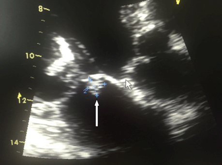
A 77-year-old male with a history of chronic obstructive pulmonary disease, mitral valve prolapse, Raynaud's phenomenon, left knee replacement, a pulmonary embolism two years ago, and chronic back pain, who underwent spinal nerve radiofrequency ablations on October 13 and on November 10, 2015. He presented to our hospital with a weight loss of 15 pounds over a period of three months, a subjective fever in the previous one month, and altered mental status for one day prior to admission. The review of systems was negative for chills, sweats, flu-like symptoms, headache, vision changes, sore throat, earache, neck pain, cough, shortness of breath, abdominal pain, nausea, vomiting, diarrhea, constipation, back or joint pain, rash, or genitourinary complaints. No injuries were reported. There was no history of antibiotic use in the past 90 days or international travel. Home medications included vitamin D 5000 units daily and acetaminophen extra strength 500 mg, two tablets every six hours as needed. He had quit smoking six months previously. There was no family history of recurrent infections. His temperature was 97.8°F, pulse 72 beats per minute, respirations 18 breaths per minute, blood pressure 102 mmHg/68 mmHg, and pulse oximetry was 97% on two liters oxygen. Patient weighs 75 kg. On physical examination, he was alert and appropriately verbally responsive. His neck was supple and no cervical lymphadenopathy was palpable. The heart examination did not reveal any murmurs, rubs, or gallops and the lungs were clear to auscultation. There was no splenomegaly or spinal tenderness. The extremities were without edema, splinter hemorrhages, Osler's nodes, or Janeway lesions. Initial labs were: white blood cell count 8,600/uL, with neutrophilia of 83.8%, hemoglobin 10.7 g/dL, and platelet 161,000/uL, sodium 129 mmol/L and creatinine 1.0 mg/dL. Erythrocyte sediment rate 105 mm/hr and C-reactive protein 8.04 mg/dL. Urinalysis was positive for large blood and 8–12 red blood cells per high powered field. Chest X-ray and CT chest without contrast were both negative for acute airspace disease and a pleural effusion. A CT scan of brain revealed severe chronic microvascular disease with age related volume loss. Computed tomography scan of lumbar spine without contrast did not show any fluid collection, but multilevel degenerative disk disease with moderate spinal canal stenosis from L1 to S1 was noted. The patient was admitted to the hospital and was started on piperacillin/tazobactam and received a one gram dose of intravenous vancomycin in the emergency room. After admission, patient did not have any documented fevers. Blood cultures obtained prior to initiation of antibiotics showed Streptococcus pseudoporcinus with confirmation tests repeated three times. Subsequently, the antibiotic was switched to ceftriaxone. A repeat blood culture was negative. Echocardiogram showed a prolapsed mitral valve and a large mobile mass on the posterior outflow valve leaflet. The mass was seen prolapsing through the coopting valve (Figure 1). Doppler showed severe mitral valve regurgitation. The left ventricular ejection fraction was 60% with normal wall motion. Patient was transferred to another hospital for valve replacement. | ||||||
| ||||||
|
Discussion
| ||||||
|
In the microbiology lab, when the patient's blood cultures became positive for gram-positive cocci in chains, a catalase test was performed. It was negative. Subsequently, the Vitek system confirmed the organism as Streptococcus pseudoporcinus. Streptococcus pseudoporcinus is a β-hemolytic Streptococcus with a wide-zone hemolysis first identified in 2006 [1]. Prior to 2006, Streptococcus pseudoporcinus was grouped under Streptococcus porcinus, which has been associated with rare cases of septicemia [2]. Most of the reported cases of Streptococcus pseudoporcinus infection were from isolates in female genitourinary tract [1] [2] [3]. Streptococcus porcinus and Streptococcus pseudoporcinus may be misidentified as Group B Streptococcus because of serological cross-reactivity [1]. In a 14-month prospective observational study done in Thailand, no isolates of Streptococcus pseudoporcinus were recovered from blood or sterile sites of any patients during their study period [4] . There was one case of a thumb infection due to Streptococcus pseudoporcinus reported in 2009 in Washington State from injury sustained by a car door jamb [5]. In this report, the authors believed that the patient acquired the infection from his wife's vaginal tract. To our knowledge, there has been no report of Streptococcus pseudoporcinus subacute endocarditis. Four out of five reported cases of bacteremia due to Streptococcus pseudoporcinus identified in a study were female while the gender of the remainder case was unidentified [6]. Streptococcus porcinus and Streptococcus pseudoporcinus are hippurate hydrolysis negative, while Streptococcus agalactiae is positive. Streptococcus porcinus is about 100% Voges-Proskauer positive while only about half of Streptococcus pseudoporcinus strains are Voges-Proskauer positive [5] [7]. Streptococcus porcinus and Streptococcus pseudoporcinus can be differentiated from Streptococcus agalactiae by fermentation of mannitol and sorbitol. However, because of the similarities in the biochemical characteristics, 16s rRNA gene sequencing is necessary to differentiate Streptococcus pseudoporcinus from Streptococcus porcinus [1] [2][3][4][5][6][7]. We report a rare case of Streptococcus pseudoporcinus bacteremia with secondary endocarditis from a non-female genitourinary source. We conducted a search of the English literature via OVID and PubMed; however, it did not yield any case reports of subacute Streptococcus pseudoporcinus endocarditis to date. | ||||||
|
Conclusion
| ||||||
|
Streptococcus pseudoporcinus is mostly associated with female genitourinary infections. No known association of Streptococcus pseudoporcinus with endocarditis has been reported previously. We have reported a rare case of Streptococcus pseudoporcinus bacteremia associated with endocarditis from a non-genitourinary source. This organism may be an emerging pathogen in the etiology of mitral valve endocarditis. | ||||||
|
Acknowledgements
| ||||||
|
We are thankful to Dr. Rameshchandra Dabhi, at Peconic Bay Medical Center-Northwell Health for the echocardiogram image. | ||||||
|
References
| ||||||
| ||||||
|
[HTML Abstract]
[PDF Full Text]
|
|
Author Contributions
Peir-Yu Fang – Substantial contributions to conception and design, Acquisition of data, Analysis and interpretation of data, Drafting the article, Revising it critically for important intellectual content, Final approval of the version to be published Sandeep A. Gandhi – Analysis and interpretation of data, Revising it critically for important intellectual content, Final approval of the version to be published |
|
Guarantor of submission
The corresponding author is the guarantor of submission. |
|
Source of support
None |
|
Conflict of interest
Authors declare no conflict of interest. |
|
Copyright
© 2017 Peir-Yu Fang et al. This article is distributed under the terms of Creative Commons Attribution License which permits unrestricted use, distribution and reproduction in any medium provided the original author(s) and original publisher are properly credited. Please see the copyright policy on the journal website for more information. |
|
|