|

|
 |
|
Case Series
| ||||||
| The distally pedicled peroneus brevis muscle and fasciocutaneous sural artery flap for reconstruction of the distal third of lower leg | ||||||
| Ingo Schmidt | ||||||
|
SRH Poliklinik Gera GmbH, Straße des Friedens 122, 07548 Gera, Germany.
| ||||||
| ||||||
|
[HTML Abstract]
[PDF Full Text]
[Print This Article]
[Similar article in Pumed] [Similar article in Google Scholar] 
|


| How to cite this article |
| Schmidt I. The distally pedicled peroneus brevis muscle and fasciocutaneous sural artery flap for reconstruction of the distal third of lower leg. Int J Case Rep Images 2017;8(1):17–21. |
|
Abstract
|
|
Introduction:
The use of distally pedicled peroneus brevis muscle and fasciocutaneous sural artery flap for coverage of the distal end of lower leg is recommended for soft tissue defects with exposure of bones and/or tendons in patients who are not willing or healthy enough to undergo free microvascular tissue transplantation, and do not require microsurgical expertise.
Case Report: A short presentation of six cases including a short review of literature will highlight current knowledge and complications of these procedures. Conclusion: The distally pedicled peroneus brevis muscle and fasciocutaneous sural artery flaps are useful for coverage of soft tissue defects of the distal third of lower leg. In our patients, the complication rate of distally pedicled neurofasciocutaneous sural artery flap is higher than the distally pedicled peroneus brevis muscle flap. | |
|
Keywords:
Distal third lower leg, Distally pedicled peroneus brevis muscle flap, Distally pedicled sural artery flap, Soft tissue defect
| |
|
Introduction
|
|
Anatomical features of the distal third of lower leg and heel like subcutaneous bone surrounded by tendons with no muscles, vessels in isolated compartments with little intercommunication between them make the coverage of the wounds in the region a challenging problem. Options for coverage of soft tissue defects are free flaps, perforator flaps, reverse flow flaps, muscle flaps, cross leg flaps, and axial pedicled fasciocutaneous flaps such as the distally pedicled sural artery flap [1] [2][3]. Quality debridement is the key to success for the healing of wounds in this region. Negative-pressure vacuum assisted closure (VAC) therapy before soft tissue coverage provides a sterile and controlled environment that can lessen the duration of wound healing, promotes better capillary circulation, and decreases the bacterial load [4]. The use of distally pedicled peroneus brevis muscle and neurofasciocutaneous sural artery flap for coverage of the distal end of lower leg is recommended for soft tissue defects with exposure of bones and/or tendons in patients who are not willing or healthy enough to undergo free microvascular tissue transplantation, and do not require microsurgical expertise. |
|
Case Series
|
|
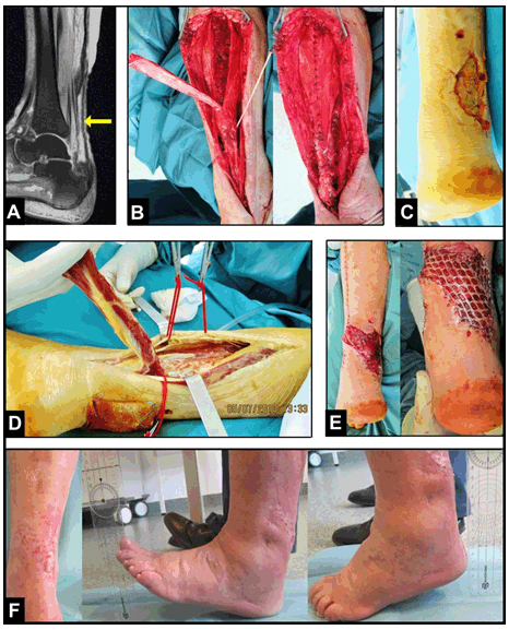
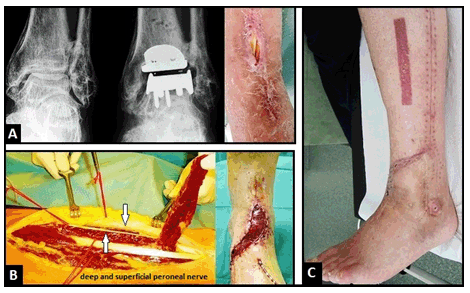
Case 1 Case 2 Case 3 Case 4 Case 5 Case 6 |
|
|
|
|
|
|
|
|
|
|
|
|
|
Discussion
|
|
Originally, the peroneus brevis was a type II muscle flap according to the classification by Mathes and Nahai [5] with a dominant pedicle from the peroneal artery which is located proximally, and distal minor pedicles from the peroneal or tibial vessels, but it was reclassified as a type IV [6]. When harvesting the muscle with the proximal segmented pedicles, it can be used as flap for coverage of the middle third of lower leg. When harvesting the distal segmented pedicles which are found within six cm from the tip of lateral malleolus (approximately three fingerbreadths), it can be used in a distally pedicled manner for the distal third of lower leg. Lorenzetti et al. reported on a flap survival of 100% of 10 patients, and the ankle functionality and stability were maintained due to preservation of peroneus longus muscle [7]. The advantage is that the donor site can always be closed primarily and the flap is relatively reliable even in high-risk patients with a number of comorbidities, but care must be taken when using this flap in patients with peripherial arterial disease [8]. The distally pedicled neurofasciocutaneous sural artery flap was first described by Masquelet et al. [2], it is a skin island flap which is retrograde supplied by at least three perforator vessels from the peroneal artery within approximately six cm from the tip of lateral malleolus. However, this flap is not free of any complications mostly based on venous congestion, and the weakness can be the pivot point. The flap's arterial inflow is robust and constant, but the venous congestion is susceptible, occurring in up to 21.4% of cases [9], and it is mostly detected if the flap was used in a 180° turned manner [10]. To prevent venous stasis intra- and early postoperatively, the pivot point of vascular pedicle including the short saphenous vein can be covered temporary with a skin substitute and covered secondary with a skin graft. Another options to prevent venous congestion are the flap's use in a two-stage manner, supercharged or superdrained manner, and/or intermittent short saphenous vein phlebotomy [10][11][12][13]. Schmidt et al. [14] reported on a survival rate of flap's use in an adipofascial manner with additional skin grafting in 87.5% of 104 cases. In cases in which the short saphenous vein cannot be found, the flap should not be utilized; and in older, high-risk, and critically multimorbid patients including peripherial arterial disease, a considerable necrosis rate of 36% of a total of 70 procedures was found by Baumeister et al. [15]. An unacceptable failure leading to a loss of flap is when the vascular pedicle was elected too short and no sufficient arterial supply exists. |
|
Conclusion
|
|
The distally pedicled peroneus brevis muscle and fasciocutaneous sural artery flaps are useful for coverage of soft tissue defects of the distal third of lower leg. In our patients, the complication rate of distally pedicled neurofasciocutaneous sural artery flap is higher than the distally pedicled peroneus brevis muscle flap. |
|
Acknowledgements
|
|
I would like to thank Henrik Eisner for his help in designing the figures. |
|
References
|
|


|
[HTML Abstract]
[PDF Full Text]
|
|
Author Contributions:
Ingo Schmidt – Substantial contributions to conception and design, Acquisition of data, Analysis and interpretation of data, Drafting the article, Revising it critically for important intellectual content, Final approval of the version to be published |
|
Guarantor of submission
The corresponding author is the guarantor of submission. |
|
Source of support
None |
|
Conflict of interest
Authors declare no conflict of interest. |
|
Copyright
© 2017 Ingo Schmidt. This article is distributed under the terms of Creative Commons Attribution License which permits unrestricted use, distribution and reproduction in any medium provided the original author(s) and original publisher are properly credited. Please see the copyright policy on the journal website for more information. |
|
|
|
About The Author
| |||
| |||