| |
|
|
|
Case Series
| ||||||
| Superficial siderosis following trauma to the cervical spine: Case series and review of literature | ||||||
| Pranab Sinha1, Sophie Jane Camp2, Harith Akram3, Robin Bhatia4, Adrian Thomas Carlos Hickman Casey5 | ||||||
|
1MRCS, BSc, MBBS, Flat 1, 28-32 London Rd, Tunbridge Wells, Kent, TN1 1BX.
2PhD, MRCS, MBBS, MA (Oxon), the National Hospital for Neurology and Neurosurgery, Queen Square, London, United Kingdom. 3FRCS (Neuro.Surg), MB ChB, the National Hospital for Neurology and Neurosurgery, Queen Square, London, United Kingdom. 4FRCS (SN), PhD, MA, MBBS, the National Hospital for Neurology and Neurosurgery, Queen Square, London, United Kingdom. 5FRCS, FRCS SW, MBBS, the National Hospital for Neurology and Neurosurgery, Queen Square, London, United Kingdom. | ||||||
| ||||||
|
[HTML Abstract]
[PDF Full Text]
[Print This Article]
[Similar article in Pumed] [Similar article in Google Scholar] 
|
| How to cite this article |
| Sinha P, Camp SJ, Akram H, Bhatia R, Casey ATCH. Superficial siderosis following trauma to the cervical spine: Case series and review of literature. Int J Case Rep Images 2017;8(1):11–16. |
|
Abstract
|
|
Superficial siderosis is a rare progressive disease associated with chronic hemosiderin deposition on the surfaces of the central nervous system (CNS). It typically manifests clinically in sensorineural hearing loss, cerebellar ataxia, and pyramidal signs. Recurrent or continuous bleeding into the cerebrospinal fluid is implicated in the disease process. The magnetic resonance imaging gradient-echo T2-weighted images have high sensitivity for hemosiderin deposits that bathe the CNS, giving the characteristic black rimmed area of hypointensity apparent on these images. The natural history and its treatments are still not clearly defined in literature. Our report details the clinical course and management of three cases of superficial siderosis following either cervical spine or brachial plexus injury. All of them underwent surgical intervention. In two of the cases, positive cessation of the intradural bleeding was achieved through surgery but clinical and radiological improvement occurred in only one of the cases. One patient had a negative intradural exploration. To date, 30 cases of superficial siderosis reported in the literature have undergone surgical intervention. Cessation of disease progression or neurological improvement has been documented in 18 of these cases. Our cases reveal that patients with superficial siderosis often develop severe functional impairment due to the progressive nature of the disease. On balance, we are of the opinion that early craniospinal imaging and surgical exploration should be undertaken, at least to attempt to halt neurological deterioration.
| |
|
Keywords:
Cervical Spine, Review, Superficial Siderosis, Trauma
| |
|
Introduction
| ||||||
|
Superficial siderosis of the central nervous system (CNS) is a rare progressive disease entity associated with chronic hemosiderin deposition in the leptomeninges and sub-pial parenchyma [1]. It typically presents with sensorineural hearing loss, cerebellar ataxia, and pyramidal signs [1] [2] [3]. Other features include cognitive impairment, bladder dysfunction, anosmia, myelopathy, back pain, bilateral sciatica, and lower motor neuron signs [4] [5] [6]. Recurrent or continuous bleeding into the cerebrospinal fluid is implicated in the aetiology of superficial siderosis. This may be as a result of intracranial or spinal neoplasm, subarachnoid hemorrhage, subdural hematoma, hemorrhagic meningitis, head injury, and spinal trauma with nerve root avulsion [3] [6]. Iatrogenic damage during surgery is a further possible cause [1] [5]. The natural history and treatment paradigms for superficial siderosis are not clearly defined. It is unknown why only a proportion of patients with hemosiderin deposition after subarachnoid hemorrhage develop the condition. Heme metabolism within the blood-brain barrier underpins the aetiology. The following report details the clinical course and subsequent management of three cases of superficial siderosis following cervical spine or brachial plexus injury. | ||||||
|
Case Series
| ||||||
|
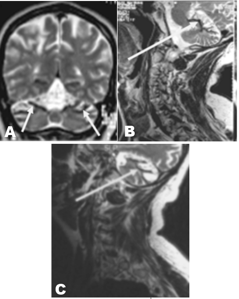
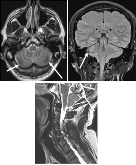
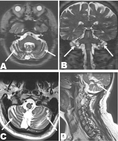
CASE 1 The patient had a broad based ataxic gait, and exhibited a cerebellar dysarthria. Examination of the cranial nerves revealed hypometric saccades, a mild left facial weakness, and a sensorineural hearing loss on the left. The intrinsic muscles of the right hand were atrophied, with clawing of the digits. Sensation was absent in the right C7, C8, and T1 dermatomes. The right triceps reflex was absent, but all other upper limb reflexes were brisk. Reflexes were slightly reduced in the lower limbs, with bilaterally down going plantars. Magnetic resonance imaging scan of brain demonstrated superficial siderosis, most markedly over the superior cerebellum, which was grossly atrophic (Figure 1A). These findings were also noted over the posterior aspect of the midbrain and around the calcarine sulci. Imaging of the cervical spine showed only degenerative changes, with a right-sided ventral longitudinal intraspinal fluid filled collections (VLISFC) at the C6/7 level. A CT myelogram to identify the dural defect was carried out and it confirmed a well-defined 20x15 mm lesion expanding into the right C7/T1 intervertebral foramen, consistent with a VLISFC. Routine blood tests revealed mild iron deficiency anemia, whilst examination of the cerebrospinal fluid (CSF) showed an increased ferritin concentration (100 IU/L). Microtrauma to the internal venous plexus in the epidural space by CSF leak may be the a reason for red blood cells seen in the cervical spine. The patient underwent a C6 to T1 laminectomies and exploration of the VLISFC in March 2008. Following a durotomy, the right C8 nerve root was inspected, and the VLISFC was also explored. There was obvious staining on inspection of the cord. A small intradural bleeding vessel was identified, this was in close proximity to the site of dural aberration but separate from dissection site. This was coagulated. The defect in the dura was then repaired. Postoperative imaging taken three months after the surgery showed that the intramedullary signal change had become less conspicuous (Figure 1B). The patient's radiological changes postoperatively were mirrored by clinical improvement in his postoperative symptoms over the course of the two years following the surgical repair. CASE 2 Brain MRI scan revealed susceptibility artefacts along the surface of the cerebellum bilaterally, which were thought to represent superficial siderosis (Figure 2). There was an appearance of a VLISFC at the level of C6 and C7. There was also an incidental finding of tonsillar ectopia in keeping with a Chiari I formation, and general non-significant cervical degenerative changes. Intracerebral and spinal angiography did not show any obvious vascular pathology. The patient's symptoms displayed signs of intracranial hypotension. There is a close association with superficial siderosis with the underlying mechanism being a dural tear [7]. Given the patient's progressive neurological deterioration, and the imaging findings consistent with superficial siderosis, the patient agreed to a surgical exploration of the VLISFC. This was undertaken in August 2008. Through a posterior spinal approach, a unilateral partial facetectomy at C6/C7 revealed the capsule of the VLISFC. The capsule was opened and the cavity fully explored using the microscope. Staining of the spinal cord was observed. No bleeding point was identified. Artificial dura and tissue glue were applied, followed by layered closure. Over the two weeks following surgery, the patient's cognitive function deteriorated. However, the patient then slowly recovered to their preoperative baseline. Subsequently, the patient experienced worsening episodes of postural headache, and their preoperative symptoms failed to improve. Postoperative imaging did not reveal a surgical complication as a cause of the headaches. Local occupational and speech and language therapists were enlisted to facilitate his activities of daily living. In addition, a local hematologist commenced deferiprone in place of trientine, to aid improvement in the patient's functional state. CASE 3 On examination the patient had anosmia, horizontal nystagmus on lateral gaze, and diplopia on right and left gaze. The patient had a complete sensorineural hearing loss on the left and partial loss on the right. The patient exhibited poor co-ordination of the right upper limb, with past pointing and dysdiadochokinesia. The tone, power, and reflexes were normal throughout all four limbs, with down going plantars. The patient walked with a wide based gait and their higher mental functions were intact. A cervical spine x-ray revealed fusion of the spinous process of C1 and C2, with interspinous wires. A cervical spine CT scan showed the wiring through the posterior elements of C1 and C2, with bony fusion. There was fusion of the peg anteriorly at C1 and fusion of C2 and C3. An MRI scan of the head revealed evidence of superficial deposition of blood degradation products primarily affecting the posterior fossa (Figure 3A). These were predominantly surrounding the midbrain, extending down the cervicomedullary junction to the superior cervical cord and lining the posterior lateral, and the fourth ventricles. Similar findings, but to a lesser degree, were seen on the surfaces of both cerebral hemispheres. There was some signal attenuation affecting the left lateral semicircular canal which was most obvious on the axial high resolution images. An MRI scan of the spine showed that the odontoid peg was malformed and posteriorly inclined. However, no thecal sac compression was reported. Metallic artefact was seen in the soft tissues posteriorly. The visualized spinal cord was normal in contour. The cervical cord also showed siderosis on its surface but otherwise was of normal signal intensity. Intracerebral and spinal angiography did not show obvious vascular pathology. This could be due to craniospinal hypovolemia, which may lead to prominent vasculature on the MRI but negative results on angiography. The patient underwent C1-C3 laminectomy through a posterior spinal approach in March, 2009. A microscopic intradural exploration revealed a bleeding point, which was separate from the dissection site. This was cauterized. The dura was then repaired. In the postoperative period, the patient noted worsening headaches, numbness of the right side of his body, and he became doubly incontinent. A subsequent MRI scan revealed a large irregular VLISFC in the posterior extraspinal tissues. The patient underwent a re-exploration of the surgical site and the VLISFC was repaired. With intense neurorehabilitation the patient regained fecal continence and the patient's urinary sphincter control improved. However, the patient's right-sided sensory loss persisted. The patient's most recent MRI showed focal cord atrophy at the level of C2. The previously noted VLISFC was no longer visualized (Figure 3B). The patient was subsequently transferred to his local neurorehabilitation unit. | ||||||
|
| ||||||
| ||||||
|
| ||||||
|
| ||||||
|
Discussion
| ||||||
|
Superficial siderosis is a rare condition [3] [8] , due to chronic hemorrhage into the subarachnoid space [1]. Historically, the diagnosis of superficial siderosis was made by histological examination of biopsied tissue, or at autopsy. However, in the modern era, MRI scan can facilitate the diagnosis, especially the gradient-echo T2-weighted images which have a high sensitivity for hemosiderin deposition [8]. Macroscopically, superficial siderosis is apparent by the brownish discoloration of the leptomeninges and adjacent parenchyma, with a predilection for the superior vermis, crest of the cerebellar folia, basal frontal lobe, temporal cortex, brainstem, spinal cord, nerve roots, and cranial nerves I, II, V, VIII, and X [6]. At a microscopic level, hemosiderin deposition occurs within macrophages in the perivascular spaces, and along the pial vessel walls. This may lead to neuronal loss, gliosis, and demyelination [6]. The hemosiderin is derived from the breakdown of heme within the glia and the microglia. These cells will synthesize ferritin, however, once their biosynthesis capabilities have been exceeded, hemosiderin is produced. Unbound ferric ions mediate apoptosis by free radical mechanisms [8]. The iron deposits on the CNS surfaces bathed by cerebrospinal fluid have a paramagnetic effect on gradient echo sequences on MRI (T2-weighted images), giving a characteristic black rimmed area of hypointensity [9] [10]. The aim of treatment is to prevent progression of the neurological deficit. Medical and surgical options have been proposed. Medical management includes the use of iron chelators, with variable success [3] [8] [11]. Trientine has been associated with iron and copper chelation, but may cause increased iron levels within the liver, presumably due to its interference with ceruloplasmin [3]. Surgical management of superficial siderosis involves identifying and terminating the source of chronic bleeding [3]. Posti et al. reported that of 27 cases undergoing surgical intervention, disease progression was halted in 13 patients, four patients clinically improved, five patients had further clinical deterioration, whilst five patients sustained other complications [12]. Kumar et al. reported a case of superficial siderosis where intradural exploration did not identified a bleeding source [7][13][14][9][10]. Egawa et al. reported two cases of superficial siderosis where the patients had dural defects with fluid-filled collections in the spinal canals, which were successfully closed [2]. One of the patients had cessation of further neurological deterioration, whilst the other partially deteriorated after surgery. To date, 30 cases of superficial siderosis reported in the literature have undergone surgical intervention, and in five individuals no source of bleeding was identified intraoperatively. Disease progression has either been halted or there has been neurological improvement in 18 of these cases thus far. All our three cases were referred late to our quaternary centre from other hospitals, this was perhaps due to delay in presentation together with difficulty in diagnosing this rare and obscure disease. All three cases had negative angiography. Previous studies of patients with cranial cerebellar superficial siderosis have shown to have negative angiography [3] [15]. The authors recognize that angiography seems to have limited sensitivity. Post-traumatic VLISFC was deemed a potential target for a bleeding vessel and therefore explored. Intraoperatively all three patients had obvious staining of the spinal cord. Authors considered biopsy of the pia arachnoid but believed it to be hazardous and of no additional benefit to the patients. The two cases which revealed the intradural microscopic bleeding vessel were entirely separate from the dissection site. The intradural spinal artery after the arachnoid layer opened is usually bloodless and were not damaged intraoperatively in these cases. The intradural microscopic bleeding vessels were likely bleeding over a period of years. This is clearly unusual and the authors cannot explain this as the normal coagulation cascade should have stopped this process. However, Tapscott el al. have previously described of a case where superficial siderosis developed more than a decade after the traumatic brachial nerve root avulsion [16]. Angiography was not able to locate a specific bleeding source but surgical repair of the meningeal diverticulum and venous cauterization helped reduce the overall central nervous system bleeding. Our cases reveal that patients with superficial siderosis often develop severe functional impairment due to the progressive nature of the disease. This may significantly compromise their quality of life. The first case showed a positive outcome at two years. However, the second case highlights the possibility of a negative intradural exploration. The third case demonstrates that quality of life is not necessarily improved even if there is confirmed cessation of an intradural bleeding point. On balance, we are of the opinion that early craniospinal imaging and surgical exploration should be undertaken, at least to attempt to halt neurological deterioration. | ||||||
|
Conclusion
| ||||||
|
Patients with superficial siderosis often develop myriad of neurological symptoms and signs that lead to severe functional impairment due to its progressive nature. On balance, we are of the opinion that early craniospinal imaging and surgical exploration should be undertaken, at least to attempt to halt neurological deterioration. | ||||||
|
References
| ||||||
| ||||||
|
[HTML Abstract]
[PDF Full Text]
|
|
Author Contributions:
Pranab Sinha – Substantial contributions to concept and design, Drafting the article, Revising it critically for important intellectual content, Final approval of the paper to be published Sophie Jane Camp – Substantial contributions to concept and design, Drafting the article, Revising it critically for important intellectual content, Final approval of the paper to be published Harith Akram – Substantial contributions to concept and design, Drafting the article, Revising it critically for important intellectual content, Final approval of the paper to be published Robin Bhatia – Substantial contributions to concept and design, Drafting the article, Revising it critically for important intellectual content, Final approval of the paper to be published Adrian Thomas Carlos Hickman Casey – Substantial contributions to concept and design, Drafting the article, Revising it critically for important intellectual content, Final approval of the paper to be published |
|
Guarantor of submission
The corresponding author is the guarantor of submission. |
|
Source of support
None |
|
Conflict of interest
Authors declare no conflict of interest. |
|
Copyright
© 2017 Pranab Sinha et al. This article is distributed under the terms of Creative Commons Attribution License which permits unrestricted use, distribution and reproduction in any medium provided the original author(s) and original publisher are properly credited. Please see the copyright policy on the journal website for more information. |
|
|