| |
|
|
|
Case Series
| ||||||
| Ultrasound imaging of tongue malignancy | ||||||
| Margaret Eseza Kisansa1, Savvas Andronikou2 | ||||||
|
1MB ChB, M Med Rad Diagnostics, Radiologist, Department of Diagnostic Radiology and Imaging, Sefako Makgatho Health Sciences University, Gauteng, South Africa.
2MBBCh, FCRad, FRCR, PhD, Radiologist, Department of Diagnostic Radiology and Imaging, Sefako Makgatho Health Sciences University, Gauteng, South Africa. | ||||||
| ||||||
|
[HTML Abstract]
[PDF Full Text]
[Print This Article]
[Similar article in Pumed] [Similar article in Google Scholar] 
|
| How to cite this article |
| Kisansa ME, Andronikou S. Ultrasound imaging of tongue malignancy. Int J Case Rep Images 2017;8(1):1–6. |
|
Abstract
|
|
Introduction:
Squamous cell carcinoma of the tongue is a common malignancy associated with risk factors like excessive alcohol consumption, heavy tobacco smoking and human papilloma virus. Magnetic resonance imaging (MRI) scan is considered to be the gold standard in investigating these tumors. However, MRI equipment is expensive to buy and is not readily available in some centers. Computed tomography scan has also been used in imaging these patients but this modality carries a radiation burden. Patient's five-year survival is dependent on early diagnosis. It is, therefore, important to diagnose early and image accurately to ensure good outcomes.
Case Series: Two male patients with confirmed carcinoma of the tongue are reported. The first patient was 65-year-old and the second patient was 40-year-old. They both presented with odynophagia. Clinical examination revealed ulcerating lesions involving the base of the tongue. Axial CT scans and ultrasound imaging were performed on these patients through the floor of the mouth. This case report focused on comparing the ability of the two modalities, in delineating margins and depicting tumor thickness. Conclusion: Ultrasound gives excellent information with regards to tumor thickness, margins and vascularity. This highlights the value of using ultrasound as an additional tool in imaging of these patients especially in regions where CT and MRI scans are not readily available. | |
|
Keywords:
Carcinoma, Tongue malignancy, Ultrasound, Ulcerating lesions
| |
|
Introduction
| ||||||
|
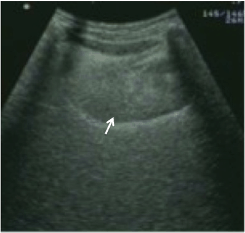
The prevalence of oral cancers is high in the world, and the risk factors cited are: excessive alcohol consumption, heavy tobacco smoking as well as human papilloma virus (HPV). Smoking tobacco and smoke less products have contributed to an increased incidence in some countries like Taiwan [1]. Computed tomography (CT) scan and magnetic resonance imaging (MRI) scan are the current modalities of choice in imaging of oral cavity tumors because of good tissue differentiation and excellent nodal mapping [2]. However, MRI machines are expensive to buy and maintain, and are not readily available in the most developing nations. Computed tomography scanners, although more available compared to MRI scanners, do use ionizing radiation which is known to carry risks. On the other hand, ultrasound machines are relatively inexpensive, readily available and easy to use. This makes a combination of ultrasound with CT, an important one in investigating patients with oral cancers in areas where MRI machines are not available. Since early detection and treatment of oral cancers is crucial for good outcomes, it makes imaging a very important component in the management of these patients. Patient's five-year survival was found to be dependent on tumor depth, with invasion greater than 2 mm being predictive of poor outcomes and a 3.7-fold increase in the risk of regional recurrence. It is therefore very crucial to measure the tumor depth accurately [3]. In this case series, imaging was done using Aloka SSD 5500 Ultrasound Unit (Aloka Japan) with a convex probe at 5.0 MHz frequency. The tongue was imaged through the sub-mandibular region (axial and sagittal planes) in these patients who had also undergone prior contrasted CT scanning of floor of the mouth. Computed tomography images were acquired on a spiral CT scanner (Spiral 4 slice Asteion CT scanner Toshiba Japan). A long axis control ultrasound of an individual with no tongue pathology is included to demonstrate the normal sonographic appearance of the tongue (Figure 1). Selected ultrasound images were compared with the corresponding axial CT images in the two patients at similar anatomic levels. | ||||||
| ||||||
|
| ||||||
|
Case Series
| ||||||
|
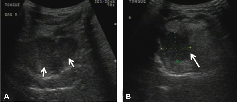
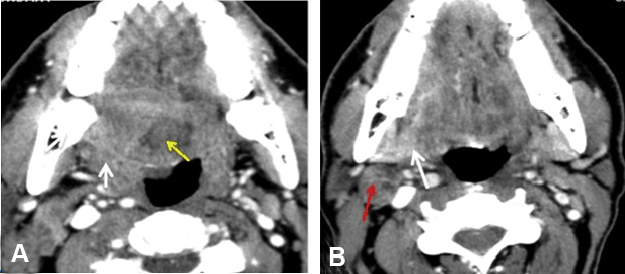
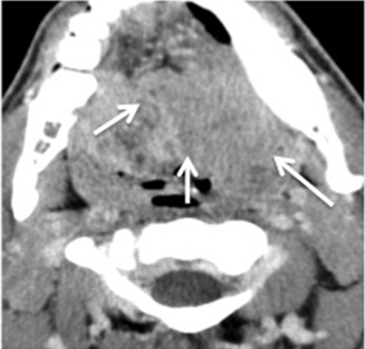
This case series presents two patients with confirmed diagnosis of carcinoma of the tongue who had undergone CT imaging both before and after intravenous contrast injection and were subsequently taken to ultrasound for further imaging. Comparisons of imaging findings, obtained from ultrasound and CT in the two patients are presented. Case 1 In comparison, the post contrast axial CT images shown in Figure 3A-B demonstrated an ill-defined hypodense lesion involving the base of the tongue on the right side. This lesion showed distortion of the oropharynx, but the margins were poorly defined. Distortion and infiltration of the right floor of the mouth and para-pharyngeal spaces was evident but the margins of the entire lesion were difficult to determine and depth of the tumor could not be measured accurately. Case 2 | ||||||
|
| ||||||
|
| ||||||
|
| ||||||
|
| ||||||
|
| ||||||
|
Discussion
| ||||||
|
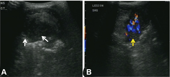
Although computed tomography (CT) and magnetic resonance imaging (MRI) scan are the modalities commonly utilized in imaging of intra-oral tumors, for accurate measurement and evaluation of the tumor margins [4], there have been some publications which clearly documented the use of intra-oral ultrasound for evaluation of these tumors. Yuen et al. showed that intra-oral ultrasonography was accurate in measuring tumor thickness and they also cited its usefulness in pre-treatment staging and prognosis [5]. A study done in Japan using intraoral ultrasound also showed that ultrasonography is an excellent imaging modality in delineating tumor margins and tumor thickness [6]. Yesuratnam et al. went further to advocate that ultrasound should be used as a first-line modality of choice for preoperative assessment of tumor thickness. This study too was based on intra-oral ultrasound imaging [7]. Doppler ultrasound has been found to be very useful in predicting grades of malignancy both in the tumor and cervical lymph nodes [8]. The sublingual approach has also been used before, with positive results. It is noted to be safe, relatively cheap and readily available [9]. In our experience, the sublingual route is comfortable, tolerable and less invasive in patients with tongue tumors, as these lesions may be ulcerated and painful. It was also found to be more tolerable for the imager, as these patients had marked halitosis. We demonstrated that ultrasound shows the primary lesion as a hypo-echoic mass with clear delineated margins and that Doppler ultrasound is useful in demonstrating the vascularity of the lesion, which aids in determining tumor staging. The tumor can be measured accurately and the tumor margins can be assessed adequately. In both the patients, CT scan failed to demonstrate the tumor edge clearly, whereas ultrasound demonstrated the tumor edge effectively. Evaluation of neck lymphadenopathy is also possible with ultrasound. | ||||||
|
Conclusion
| ||||||
|
This case series highlights the importance of ultrasound as a modality that should be embraced and used more frequently in imaging of intraoral tumors than the presented case. We are only recommending adding ultrasound to the protocol of imaging and not replacing any of the gold standard modalities MRI and CT scans. There are no reports comparing the use of sub-lingual ultrasound and CT scan in imaging of tongue malignancies and this is an area that needs further investigation. | ||||||
|
Acknowledgements
| ||||||
|
We would like to express our sincere thanks Moseley Kaboyo Kyamulesire, Paul Mbatya and Ram Kyamulesire for administrative support. | ||||||
|
References
| ||||||
| ||||||
|
[HTML Abstract]
[PDF Full Text]
|
|
Author Contributions:
Kisansa Margaret Eseza – Substantial contributions to conception and design, Acquisition of data, Analysis and interpretation of data, Drafting the article, Revising it critically for important intellectual content, Final approval of the version to be published Andronikou Savvas – Analysis and interpretation of data, Revising it critically for important intellectual content, Final approval of the version to be published |
|
Guarantor of submission
The corresponding author is the guarantor of submission. |
|
Source of support
None |
|
Conflict of interest
Authors declare no conflict of interest. |
|
Copyright
© 2017 Kisansa Margaret Eseza et al. This article is distributed under the terms of Creative Commons Attribution License which permits unrestricted use, distribution and reproduction in any medium provided the original author(s) and original publisher are properly credited. Please see the copyright policy on the journal website for more information. |
|
|
|
About The Authors
| |||
| |||
| |||