| |
|
|
|
Case Report
| ||||||
| A fever of unknown origin as a presenting symptom in toxoplasmosis: Back to earth | ||||||
| Jill F. Mentink1, Michiel van Rijn2, Adriaan Dees3 | ||||||
|
1MD, Resident, Department of Internal Medicine, Ikazia Hospital, Rotterdam, the Netherlands.
2MD, PhD, Microbiologist, Department of Microbiology, Ikazia Hospital, Rotterdam, the Netherlands. 3MD, PhD, Internist, Department of Internal Medicine, Ikazia Hospital, Rotterdam, the Netherlands. | ||||||
| ||||||
|
[HTML Abstract]
[PDF Full Text]
[Print This Article]
[Similar article in Pumed] [Similar article in Google Scholar] 
|
| How to cite this article |
| Mentink JF, van Rijn M, Dees A. A fever of unknown origin as a presenting symptom in toxoplasmosis: Back to earth. Int J Case Rep Images 2017;8(1):73–76. |
|
Abstract
|
|
Introduction:
Fever of unknown origin (FUO) and weakness in the upper legs are among the presenting symptoms in acute toxoplasmosis, which can occur in immune-competent individuals.
Case Report: A 76-year-old Caucasian male presented at the hospital with relapsing fever and weakness in both upper legs. A neurological examination confirmed weakness in the quadriceps muscles, which appeared to be atypical for any neurological substrate. An extensive work-up did not lead to a diagnosis. After two weeks of clinical investigations and persistent fever, we re-examined our patient to look for anamnestic signs. We again asked about (recent) vacations, hobbies and potential exposures. Ultimately, the patient told us he performed charity work at a care farm for more than ten years. He had contact with sheep and soil, which occurred while working in the garden without wearing gloves. A diagnosis of a primo toxoplasmosis infecton was made and the patient was treated with daraprim and clindamycin. Within two days his temperature normalized and the strength in his legs improved gradually. He achieved a complete recovery within three months. Afterwards his wife mentioned that her husband was afraid of losing his volunteer job, when telling his doctors about his work with disabled people. Conclusion: In patients presenting with FUO, clinicians in search of a diagnosis must struggle through a long list of possible diagnosis. Diagnostic delay can be shortened by considering the potential clues in the patient's history. | |
|
Keywords:
Diagnostic delay, Fever of unknown origin (FUO), Toxoplasma gondii
| |
|
Introduction
| ||||||
|
Fever of unknown origin (FUO) and weakness in the upper legs are among the presenting symptoms in acute toxoplasmosis, which can occur in immune-competent individuals. The prevalence of the obligate, intracellular protozoan Toxoplasma gondii varies worldwide, but it is generally assumed that the global infection rate is 25–30%. Some healthy individuals who are infected become symptomatic. Immunocompromised patients are at risk of reactivation of a primo infection and developing a severe and complicated illness. In patients presenting with FUO, clinicians in search of a diagnosis must struggle through a long list of possible diagnosis. In this modern era of sophisticated imaging techniques, the temptation to fully rely upon laboratory tests results and scans should be resisted [1][2]. This diagnostic delay can be shortened by considering the potentially diagnostic clues in the patient's history. | ||||||
|
Case Report
| ||||||
|
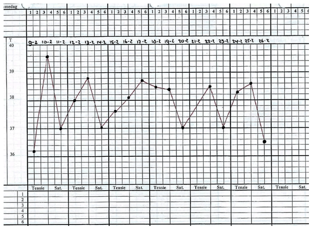
A 76-year-old male was admitted to hospital with relapsing fever along with weakness and pain in his upper legs. He said told that the weakness, which had a progressive course, had begun several weeks earlier and had progressively worsened. His medical history included diabetes mellitus type 2, hypertension, uncomplicated coronary angiographic bypass graft and laparoscopically performed cholecystectomy. Moreover, 24 years earlier, he had undergone allogeneic bone marrow transplantation because of paroxysmal nocturnal hemoglobinuria (PNH). His daily medication regime included metformin, enalapril, fluvastatin and acetylsalicylic acid. On admission the patient was in a poor condition. He had lost 4 kg, was anorectic and had a temperature of 39.0°C. On physical examination, his heart and lungs were normal. Neurologic examination: the patient was alert, febrile, Glasgow Coma Scale E4M6V5. Investigation of the lower extremities: the patient was unable to lift his feet, the sensibility was normal. Mild weakness was observed. Knee and Achilles reflexes were 0/0 and -4/-2 respectively. Laboratory tests on admission: leucocytosis of 15.9x10⁹/L, sedimentation rate in the first hour of 90 and C-reactive protein of 386 mg/L. Blood and urine samples were obtained for cultures, full blood count and chemistry, along with serological tests for underlying infectious causes and rheumatic diseases. A chest X-ray and abdominal ultrasound were performed. A presumptive diagnosis of infection was made and because of the lack of focus we decided not to start antibiotics but wait for the results of the cultures. A neurologist was consulted, but found no central or peripheral abnormalities, apart from painful quadriceps muscles, that might explain the clinical picture. We did not perform a muscle biopsy at that moment. During his stay in the hospital, a relapsing fever was observed, that met the criteria of FUO, also known as febris e causa ignota (Figure 1). Cultures of blood and urine remained negative. Autoimmune serology and ANCA's were absent. Based on serology hepatitis, HIV, Borrelia, influenza, syphilis, Coxiella burnetii (Q-fever) and brucellosis were ruled out. The serological tests for Epstein-Barr and cytomegalic virus were positive for past infections. Withdrawal of fluvastatin did not result in any improvement. A thoracic and abdominal computer tomography (CT) scan did not reveal solid masses, abscesses, lymphadenopathy or any sign of malignancy, except for a benign cyst in the liver. A whole body (18)F-fluorodeoxyglucose ((18)F-FDG) position emission tomography (PET)-CT scan showed some, perhaps physiological uptake in the kidneys and bowel. Bone marrow aspiration and immunology analyses revealed normal cell lines following the previous transplantation, without signs of leukaemia or myeloproliferative disease. Throughout this diagnostic process, we failed to arrive at a definite diagnosis. The patient remained febrile and his condition progressively worsened in a couple of days. We decided to return to the patient to seek out potentially diagnostic clues that we had previously neglected. Subsequently, the patient mentioned that he had travelled to Brazil several years ago, but was not sure whether he had received all the proper vaccinations. Thereafter, a thick blood smear was performed to exclude malaria parasites, as well as tests for schistosomiasis and Q-fever. Eventually, the patient told us that he had worked as a charity worker for more than ten years at a care farm for disabled people. He had contact with sheep and also maintained the gardens without wearing gloves while working with soil. Afterwards his wife told us that he had been very afraid to lose his volunteer job, as a consequence of telling his doctors this information. Finally, we found that toxoplasmosis serology for IgM and IgG were positive with a rather low avidity of 0.548, which fit the diagnosis of acute infection. We considered this a primo infection in an immunocompetent host. We started our patient on daraprim (pyrimethamine) and clindamycin. Daraprim is a folic acid antagonist that is highly effective against toxoplasma especially when used in combination with sulphonamide. We preferred the combination with clindamycin because of less symptoms and similar effectiveness. We started with one dose of 100 mg Daraprim and continued with 50 mg once daily in combination with clindamycin 600 mg four times daily for two weeks. The patient was regularly seen at outpatient clinic after his discharge from hospital,. His condition gradually improved, without a return of complaints or fever. The patient remained seropositive (IgM and IgG) during six months of follow-up. | ||||||
| ||||||
|
Discussion
| ||||||
|
This report illustrates the pitfalls in the diagnostic route of a patient, who may present with a fever of unknown origin. During the ward grand rounds we discussed the immune status of this patient. He was known to have diabetes type two. He exhibited no previous signs of recurrent infections. He had undergone bone marrow transplantation because of a benign disease more than 20 years ago. However, after we made the diagnosis, we wondered whether our initial assumption was right. Very few healthy individuals who have been infected with Toxoplasma gondii become symptomatic, although fever and myalgia have been described as presenting symptoms, even in immunocompetent individuals [3][4]. The IgG and IgM antibodies were positive at presentation, with a low avidity of 0.548 for IgG. The IgM antibodies usually present within the first week of presentation, peak in one month, but may persist for a year or more. The IgG titer helps to distinguish an acute infection from a past infection [5]. A low avidity is associated with an acute infection, a high avidity makes an past infection or re-activation more likely. Toxoplasma gondii is an obligate intracellular protozoan. The prevalence of this single cell parasite differs worldwide, but is generally assumed that that the infection rate of the global population is 25–30% [6]. A high prevalence infection occurs in tropical Africa and Latin America. A total of three major genotypes have been identified, among which type I is the most virulent. Cats facilitate the sexual parasitic cycle and spreading of oocysts though faeces. Humans can acquire Toxoplasma by eating undercooked meat, drinking contaminated water, swallowing parasites after contact with cat feces (via contaminated soil or cleaning a cat litter box) or they are infected by receiving an infected organ transplant or donor blood. Some infected individuals experience non-specific flu-like symptoms. By contrast, immunocompromised patients are at risk of developing severe toxoplasmosis infections after primary infection or re-activation that may result in myocarditis, pneumonitis, polymyositis, meningoencephalitis or other conditions. Morbidity is even higher after infection with a type one or atypical strain. Afterwards we asked the transplantation centre that had previously treated the patient, whether his toxoplasmosis serology had been noted in the charts prior to transplantation. This information could not be located. The role of the FDG-PET/CT scan may be questioned. This nuclear imaging test has shown its utility in patients with FUO, along with reasonable performance [7]. In the patient presented here, the diagnostic yield of CT and PET was unclear. In contrast to the imaging techniques, our search for specific diseases based on clinical reasoning and serology was more successful [8]. | ||||||
|
Conclusion
| ||||||
|
The case demonstrates that FUO and weakness in the upper legs can be the presenting symptoms in toxoplasmosis and that this might present in immune-competent individuals. Unravelling the patient history can shorten the diagnostic delay. The diagnostic clue in this case turned out to be the patient's previous social work on a charity farm. The clinical course of the patient suggested an acute infection, however a reactivation from a prior infection, in case of a transplantation history, could not be excluded definitely. | ||||||
|
References
| ||||||
| ||||||
|
[HTML Abstract]
[PDF Full Text]
|
|
Author Contributions
Jill F. Mentink – Substantial contributions to conception and design, Acquisition of data, Analysis and interpretation of data, Drafting the article, Revising it critically for important intellectual content, Final approval of the version to be published Michiel van Rijn – Analysis and interpretation of data, Drafting the article, Revising it critically for important intellectual content, Final approval of the version to be published Adriaan Dees – Substantial contributions to conception and design, Acquisition of data, Analysis and interpretation of data, Drafting the article, Revising it critically for important intellectual content, Final approval of the version to be published |
|
Guarantor of submission
The corresponding author is the guarantor of submission. |
|
Source of support
None |
|
Conflict of interest
Authors declare no conflict of interest. |
|
Copyright
© 2017 Jill F. Mentink et al. This article is distributed under the terms of Creative Commons Attribution License which permits unrestricted use, distribution and reproduction in any medium provided the original author(s) and original publisher are properly credited. Please see the copyright policy on the journal website for more information. |
|
|