
  |

|
 |
|
Case Report
| ||||||
| Erythema ab igne in patients with dementia: Implications for caregivers | ||||||
| Zijian Zheng1, Sid Danesh2 | ||||||
|
1Medical student, Des Moines University College of Osteopathic Medicine, Des Moines, IA50312 USA.
2MD, Associate professor, Department of Dermatology, University of Southern, California, Los Angeles, CA91776 USA. | ||||||
| ||||||
|
[HTML Abstract]
[PDF Full Text]
[Print This Article]
[Similar article in Pumed] [Similar article in Google Scholar] 
|


| How to cite this article |
| Zheng Z, Danesh S. Erythema ab igne in patients with dementia: Implications for caregivers. Int J Case Rep Images 2017;8(1):70–72. |
|
Abstract
|
|
Introduction:
Erythema ab igne is a benign, asymptomatic skin condition caused by heat exposure. It is an uncommon condition, making it challenging for physicians and healthcare providers to diagnose and follow-up for potential progression to malignancy, especially in patients with dementia.
Case Report: Herein, we present a case of erythema ab igne in a 70-year-old male with dementia, in whom careful history taking from family was required to elicit the cause (a warming blanket) and diagnosis. Conclusion: Erythema ab igne can be recognized and prevented if physicians have a high index of suspicion and are aware of the history and signs. Patients with dementia require particularly close clinical follow-up. | |
|
Keywords:
Dementia, Erythema ab igne, Squamous cell carcinoma
| |
|
Introduction
| ||||||
|
Erythema ab igne is a benign cutaneous disease caused by prolonged heat exposure [1]. Although uncommon, dementia patients are particularly susceptible due to lack of self-care and cognitive impairment. Furthermore, they find the skin condition difficult to monitor, particularly for secondary malignant transformation. Here we report a case of erythema ab igne in an elderly male with dementia to illustrate that a high index of suspicion, close observation, and prompt treatment are required in this vulnerable population. | ||||||
|
Case Report
| ||||||
|
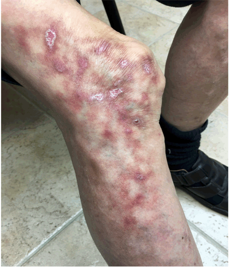
A primary care physician (PCP) referred a 70-year-old Asian male with a history of dementia, chronic heart failure, and back pain to the dermatology clinic due to a three-month history of unexplained leg rash. Most history was obtained via family members due to the patient's dementia, who reported multiple scaly rounds to oval shaped blisters on the lower limbs. On examination, the patient was alert and not in distress. He was oriented in person, time, and place and vital signs were stable and within normal range. The patient had general dry and loose skin due to aging with multiple verrucous, hyperkeratotic lesions. There were multiple varicose veins on the posterior lower legs and benign nevi on the face, upper trunk, and abdomen. The lesion of interest was a non-tender violaceous, macular rash resembling livedo reticularis on the right lower lateral thigh and upper lateral leg (Figure 1). The lesion blanched with alternating red pigmentation and normal skin. Healing wounds and hyperkeratotic scars were also present. There were no joint effusions or warmth in the lower extremity joints, which had a full range of both active and passive movement. Motor and sensory examinations were normal. Although the patient had difficulty distinguishing present and past events, he reported no pruritus, pain, or bleeding. His family reported no history of bleeding disorders, anticoagulant use, or similar past events. Apart from 81 mg aspirin every other day, he was not on any other prescribed antiplatelet or anticoagulant therapy. The patient's PCP had recently performed a well adult check that revealed normal hematological and biochemical parameters. Further direct questioning of the patient and family revealed that patient had lower back pain and that he was reluctant to take pain medication. He had instead been using an electrical warming blanket to alleviate his back pain. His family often found him forgetting to turn it off and sleeping with it in place. The patient lay on his left side at sleep. Given the history and findings, erythema ab igne was diagnosed. The patient was given triamcinolone cream and the lesions had resolved by the next visit. Instructions on prevention of similar symptoms were given to patient and the caregiver. | ||||||
| ||||||
|
Discussion
| ||||||
|
Erythema ab igne describes persistent, blanching erythematous areas that gradually develop into reticulated pigmentation and focal epidermal atrophy due to excessive or long-term heat exposure. The heat is not usually hot enough to cause overt burning in most cases. Hemostasis cause by the heat leads to a mottled appearance and subsequent pigmentation. The pigmentation can be erythematous, violaceous, pink, gold or dark brown. Various heat sources have been reported to cause erythema ab igne including heating pads, heating pans, and even laptops [2]. In our patient, a heating blanket led to his symptoms and signs. In most cases, after removal of the heating source, pigmentation gradually resolves but permanent pigmentation is possible [3]. A skin biopsy should be obtained to rule out other etiologies rather than to confirm the clinical diagnosis of erythema ab igne, which has non-specific histopathologic findings [4]; indeed, the biopsy taken in this case showed only a sparse perivascular infiltrate and non-specific findings. In early disease, epidermal atrophy, lymphocytic infiltrates, and epithelial atypia may be seen with dermal elastin deposition seen in late disease. Epithelial atypia can progress into squamous cell carcinoma, which is preventable with 5-fluorouracil or imiquimod. The use of steroid cream may help to reduce the pigmentation [5]. Livedo reticularis and other vasculitides should form part of the differential diagnosis, but our patient's recent normal laboratory results helped us to rule out any systemic vasculitis. Further, the patient did not have other symptoms such as fever or weight loss. Close follow-up of patients with erythema ab igne is crucial, not only due to a risk of malignancy but also because it is hard for patients to monitor skin changes on the lower back and at other difficult to see locations. Ulceration and worsening of the lesion warrant biopsy to rule out squamous cell and Merkel cell carcinomas, both of which have been described in association with the condition [6]. Dementia patients in particular need close follow-up since they are less likely to self-monitor and may continue to apply heat. Physicians and other healthcare providers managing dementia patients need to pay close attention to the use of heating sources, and make sure that they are properly used, and need to be aware of the cutaneous manifestations of benign and malignant diseases arising from heat exposure to ensure prompt diagnosis and management. | ||||||
|
Conclusion
| ||||||
|
Erythema ab igne can be recognized and prevented if physicians have a high index of suspicion and are aware of the history and signs. Patients with dementia require particularly close clinical follow-up. | ||||||
|
References
| ||||||
| ||||||

|
[HTML Abstract]
[PDF Full Text]
|
|
Author Contributions
Zijian Zheng – Substantial contribution to the concept and design, Drafting the article, Final approval of the version to be published Sid Danesh – Substantial contribution to the concept and design, Drafting the article, Final approval of the version to be published |
|
Guarantor of submission
The corresponding author is the guarantor of submission. |
|
Source of support
None |
|
Conflict of interest
Authors declare no conflict of interest. |
|
Copyright
© 2017 Zijian Zheng et al. This article is distributed under the terms of Creative Commons Attribution License which permits unrestricted use, distribution and reproduction in any medium provided the original author(s) and original publisher are properly credited. Please see the copyright policy on the journal website for more information. |
|
|