
  |

|
 |
|
Case Report
| ||||||
| Myxoedema: A rare cause of massive ascites | ||||||
| Rabab Fouad1, Mohamed B. Hashem1, Mohamed Said1, Marwa Khairy1, Mahmoud Abouelkhair1, A. A. Helmy1 | ||||||
|
1Endemic Medicine and Hepatogastroenterology Department, Faculty of Medicine, Cairo University, Cairo, Egypt.
| ||||||
| ||||||
|
[HTML Abstract]
[PDF Full Text]
[Print This Article]
[Similar article in Pumed] [Similar article in Google Scholar] 
|


| How to cite this article |
| Fouad R, Hashem MB, Said M, Khairy M, Abouelkhair M, Helmy AA. Myxoedema: A rare cause of massive ascites. Int J Case Rep Images 2017;8(1):66–69. |
|
Abstract
|
|
Myxoedema ascites is a rare condition with a known incidence of 4% which makes it in the bottom of the list of causes of ascites, that is why diagnosis is often delayed and physicians usually do not put it in the preliminary differential diagnosis of a case of ascites. We here report a case of Ascites due to hypothyroidism which markedly improved after thyroxin therapy. To our knowledge, this makes it the first case reported in Egypt with myxoedema ascites. Myxoedema ascites is characterized by the dramatic response to replacement therapy. The message to be taken is that myxoedema is a rare cause of massive ascites but should be evaluated if suspected since the condition is easily controlled by medical treatment.
| |
|
Keywords:
Massive ascites, Myxoedema, Serum ascites albumin gradient (SAAG)
| |
|
Introduction
| ||||||
|
Portal hypertension secondary to liver cirrhosis is the leading cause of ascites (more than 80% of cases) and peritoneal involvement in patients with malignant diseases is the second at about 10% [1]. However, some cases maybe due to other rare diseases including hypothyroidism which is characterized by a marvelous response to treatment. Patients with ascites due to portal hypertension and nephrotic syndrome usually respond to diuretic therapy while patients with ascites due to other disorders whether biliary, pancreatic, myxoedema etc. respond to treatment of the underlying cause. That is why it is essential to search for the underlying etiology of ascites carefully. | ||||||
|
Case Report
| ||||||
|
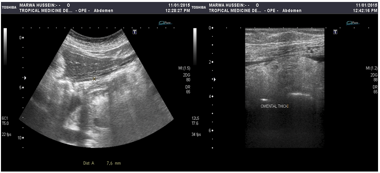
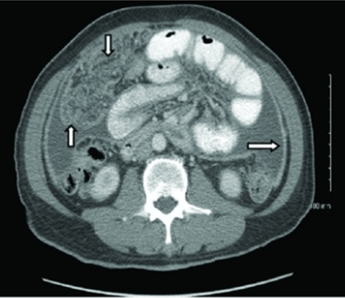
A 48-year-old female was presented with one year history of progressive abdominal distension, bilateral lower limb edema and chronic constipation. On admission, her blood pressure was 120/70 mmHg, pulse rate was 70 bpm and BMI was 26 kg/m2. General examination as well as cardio-respiratory examination were free apart from bilateral partially pitting lower limb edema reaching the thighs, with overlying slightly rough skin. The abdomen was markedly distended, with tight skin and liver was mildly enlarged. There was also shifting dullness detected by percussion. Urine analysis was normal with no evidence of proteinuria, complete blood count showed mild normocytic normochromic anemia with hemoglobin of 10.3 g/dL while leukocyte and platelet counts were normal. Regarding liver biochemical profile, albumin was 2.9 (normal range 3.5–5.5), total proteins 6.2 (normal range 6.5–8.5), total bilirubin 0.3 (normal range 0.2–1.2), ALT 23 (normal range 12–37), AST 30 (normal range 25–65), ALP 70 (normal range 50–130), GGT 26 (normal range up to 42), PC 88% (normal range 70–130), INR 1.07. Renal functions, electrolytes, HBsAg and HCV Ab were all negative. Ascitic fluid analysis was done next showing clear sample containing total leukocyte count of 200 cells of mixed cellularity and negative for malignant cells. Total proteins were 4.2 and serum-albumin ascitic gradient (SAAG) was 0.9. Gram staining, bacterial, fungal and MGIT culture (Mycobacterial growth indicator tube) cultures were all negative. Echocardiography was done which ruled out congestive heart failure as a cardiogenic causes of Ascites showing dilated left atrium, dilated left ventricle, left ventricular grade I diastolic dysfunction with an ejection fraction of 42%. We performed imaging studies to evaluate the possible cause of the ascites, Ultrasonography (Figure 1A-B) and computed tomography (CT) scan (Figure 2) of the abdomen revealed bright hepatomegaly, massive ascites, parietal and visceral peritoneal thickening as well as an echogenic omental thickening seen in the mid line raising the possibility of local peritoneal disease. The findings yielded from the imaging studies, we decided to rule out the possible causes of peritoneal diseases including peritoneal malignancies and tuberculosis so tuberculin skin test, chest X-ray and tumor markers were done showing no abnormalities. The next crucial step was to perform ultrasound guided biopsy of the thickened heterogeneous omentum where the specimen revealed moderate inflammatory reaction with possible fat necrosis, no granulomas and no malignancy encountered. At this stage, we started evaluation of other unusual causes of high protein content with low SAAG ascites so complete thyroid profile was done showing TSH 25.40 ulU/ml (normal range 0.4–4 ulU/ml), T3 < 0.3 pg/ml (normal range 2–4.4 pg/ml) and T4 0.1 ng/dl (normal range 0.8–1.9 ng/dl). Thyroid ultrasound was done next showing no abnormality in both lobes. Thyroxin was started with gradually increasing doses of levothyroxine, from 0.05 mg to 0.12 mg daily to treat the patient's hypothyroidism. However, after one month of treatment there was no improvement in her condition despite being euthyroid. This forced us to re-evaluate the diagnosis since the incidence of ascites in myxoedema is rare (4%). Also, there was omental and peritoneal thickening which are strongly suggestive of the presence of local peritoneal diseases. Furthermore, there was no accepted improvement in the amount of ascites after one month of treatment. A laparoscopy was done showing omental, visceral and parietal peritoneal thickening which were biopsied and their histopathological examination showed fibro fatty tissue infiltrated by chronic inflammatory cells with fat necrosis. That was consistent with the previous biopsy taken under ultrasound guidance so the patient was discharged after increasing the dose of levothyroxine to 0.15 mg and followed-up over the next three months where follow-up showed dramatic improvement in her condition; the ascites resolved completely and did not recur and thyroid function tests were maintained. The latest thyroid profile showed TSH 3.0 ulU/ml, T3 3.6 pg/ml and T4 2.1 ng/dl. | ||||||
| ||||||
|
| ||||||
|
Discussion
| ||||||
|
Most patients with ascites usually suffer from liver cirrhosis [2]. In about 15% of patients with ascites, there is a non-hepatic cause of fluid retention. Successful treatment is dependent on an accurate diagnosis of the cause of ascites; e.g., peritoneal carcinomatosis does not respond to diuretic therapy. Patients with ascites should be questioned about risk factors for liver disease [3]. Past history of cancer, heart failure, renal disease, thyroid disease or tuberculosis is also relevant. Hemophagocytic syndrome can masquerade as cirrhosis with ascites. These patients have fever, jaundice, and hepatosplenomegaly, usually in the setting of lymphoma or leukemia [4]. The serum ascites albumin gradient (SAAG) is formula used to assist in determining the etiology of ascites. The SAAG is the best single test for classifying ascites into portal hypertensive (SAAG >1.1 g/dL) and non-portal hypertensive (SAAG < 1.1 g/dL) causes. Calculated by subtracting the ascitic fluid albumin value from the serum albumin value, it correlates directly with portal pressure. The specimens should be obtained relatively simultaneously. The accuracy of the SAAG results is approximately 97% in classifying ascites. The terms high-albumin gradient and low-albumin gradient should replace the terms transudative and exudative in the description of ascites. Hypothyroidism though being relatively common condition yet, it manifests with ascites in a minority of patients [5]. The SAAG is usually low in cases of ascites caused by myxoedema [6]. The mechanisms by which a patient with myxoedema develops ascites is unknown. There have, however, been several hypotheses proposed. One of them suggested that ascites occurs due to the extravasation of plasma proteins as a result of abnormal capillary permeability. Another theory showed that accumulation of hyaluronic acid under the skin of patients with myxoedema may lead to ascites by a direct hygroscopic effect. | ||||||
|
Conclusion
| ||||||
|
To conclude, the message to be delivered here is that myxoedema though being a rare cause of ascites, should be excluded early especially in cases with vague presentation since it carries an excellent prognosis with adequate replacement therapy. | ||||||
|
Acknowledgements
| ||||||
|
We would like to express our deep gratitude to Professor Iman Hamza, Professor Waleed Fouad, Professor Mohamed Seif, Dr. Ismail Anwar, Dr. Reham Abd El Moniem, Dr. Eman Adel, Dr. Hedy Ayman and Dr. Yasmine Gaber for their everlasting efforts and contribution in diagnosing and treating this case. | ||||||
|
References
| ||||||
| ||||||

|
[HTML Abstract]
[PDF Full Text]
|
|
Author Contributions
Rabab Fouad – Substantial contributions to conception and design, Acquisition of data, Analysis and interpretation of data, Drafting the article, Revising it critically for important intellectual content, Final approval of the version to be published Mohamed B. Hashem – Substantial contributions to conception and design, Acquisition of data, Analysis and interpretation of data, Drafting the article, Revising it critically for important intellectual content, Final approval of the version to be published Mohamed Said – Substantial contributions to conception and design, Acquisition of data, Analysis and interpretation of data, Drafting the article, Revising it critically for important intellectual content, Final approval of the version to be published Marwa Khairy – Substantial contributions to conception and design, Acquisition of data, Analysis and interpretation of data, Drafting the article, Revising it critically for important intellectual content, Final approval of the version to be published Mahmoud Abouelkhair – Substantial contributions to conception and design, Acquisition of data, Analysis and interpretation of data, Drafting the article, Revising it critically for important intellectual content, Final approval of the version to be published A. A. Helmy – Substantial contributions to conception and design, Acquisition of data, Analysis and interpretation of data, Drafting the article, Revising it critically for important intellectual content, Final approval of the version to be published |
|
Guarantor of submission
The corresponding author is the guarantor of submission. |
|
Source of support
None |
|
Conflict of interest
Authors declare no conflict of interest. |
|
Copyright
© 2017 Rabab Fouad et al. This article is distributed under the terms of Creative Commons Attribution License which permits unrestricted use, distribution and reproduction in any medium provided the original author(s) and original publisher are properly credited. Please see the copyright policy on the journal website for more information. |
|
|