
  |

|
 |
|
Case Report
| ||||||
| Escherichia coli sepsis and pyomyositis following allogeneic stem cell transplant | ||||||
| Folusakin Ayoade1, Mohammed Alam2, Amy Bozeman3, Breanne Peyton-Thomas4, Richard Mansour5, Nebu Koshy6 | ||||||
|
1MD, Folusakin Ayoade, Clincal Fellow, Department of Medicine, Louisiana State University Health Science Center, Shreveport, LA 71103, USA.
2MD, Mohammed Alam, Assistant Professor, Department of Medicine, Louisiana State University Health Science Center, Shreveport, LA 71103, USA. 3PA, Amy Bozeman, Physician Assistant, Bone Marrow Transplant Unit, University Health, Shreveport, LA 71103, USA. 4Pharm D, Breanne Peyton-Thomas, Clinical Pharmacist, University Health, Shreveport, LA 71103, USA. 5MD, Richard Mansour, Associate Professor, Department of Medicine, Louisiana State University Health Science Center, Shreveport, LA 71103, USA. 6MD, Nebu Koshy, Associate Professor, Department of Medicine, Louisiana State University Health Science Center, Shreveport, LA 71103, USA. | ||||||
| ||||||
|
[HTML Abstract]
[PDF Full Text]
[Print This Article]
[Similar article in Pumed] [Similar article in Google Scholar] 
|


| How to cite this article |
| Ayoade F, Alam M, Bozeman A, Peyton-Thomas B, Mansour R, Koshy N. Escherichia coli sepsis and pyomyositis following allogeneic stem cell transplant. Int J Case Rep Images 2017;8(1):57–61. |
|
Abstract
|
|
Introduction:
Manifestation of infections in hematopoietic stem cell transplant patients tend to be subtle, with subsequent delay in diagnosis and effective therapy. Traditionally, in both normal and immunocompromised hosts, pyomyositis is often attributable to gram positive pathogens such as Staphylococcus aureus. Pyomyositis due to Escherichia coli is quite rare in the hematopoietic stem cell transplant host, often with uncharacteristic or atypical presentations.
Case Report: A 53-year-old male with relapsed acute myelogenous leukemia received allogeneic stem cell transplant. After eight days post-transplantation, he developed fever and other non-specific symptoms, followed by progressive left calf swelling and pain. The first two ultrasound studies of the affected leg showed no drainable collections despite obvious signs of local infection. The third ultrasound study, done after seven days of onset of symptoms, however, revealed a developing abscess with extensive surrounding cellulitis. Lower extremity computed tomography scan confirmed a 3.4x2.5 cm lesion with a central necrotic portion measuring approximately 1.8x1 cm within the lateral head of the gastrocnemius muscle. Blood cultures and drained pus from the affected calf muscle grew quinolone-resistant Escherichia coli which was sensitive to beta-lactamase antibiotics. Successful treatment was accomplished with three weeks of intravenous meropenem and abscess drainage. Conclusion: This case illustrates the unique peculiarities of infection manifestations in the immunocompromised host, especially recipients of stem cell transplant. The causative pathogen could be atypical, and the clinical and expected imaging findings may be delayed or even absent. | |
|
Keywords:
Abscess, Escherichia coli, Hematopoietic stem cell transplant, Pyomyositis, Quinolone
| |
|
Introduction
| ||||||
|
Hematopoietic stem cell transplant patients are a unique population and clinical manifestations of infection in this group are often subtle, with subsequent delay in diagnosis and effective therapy. Traditionally, most cases of pyomyositis are attributable to Staphylococcus aureus in both normal and immunocompromised hosts [1]. Despite this observation, pyomyositis in the transplant patient and similar immunocompromised hosts often have uncharacteristic presentations and are sometimes associated with the 'unlikely' pathogen [2]. | ||||||
|
Case Report
| ||||||
|
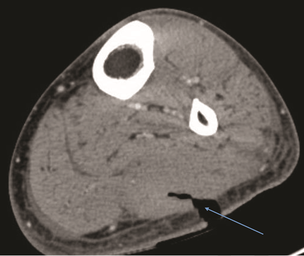
A 53-year-old white male with relapsed acute myelogenous leukemia was admitted to our hospital for allogeneic stem cell transplant. Conditioning regimen was fludarabine and melphalan and he received prophylactic levofloxacin 500 mg orally daily starting day-7 post-transplant. On day-8 post-transplant, he developed low grade fever with chills, headache, nausea, vomiting, diarrhea, decreased appetite and myalgia. The patient's vital signs were temperature 100.6°F (Tmax 100.9 after 24 hours), heart rate 112 beats/minute, respiratory rate 20 cycles/minute and blood pressure 95/63 mmHg (down from 154/87 mmHg 12 hours earlier). Physical examination findings at the time of fever including neck, lungs, heart, abdomen and musculoskeletal examinations were all within normal limits. He had a tri-fusion catheter on right chest wall with no evidence of surrounding erythema or tenderness. Before the onset of fever, he had been severely neutropenic for several days (total white blood cell in blood less than 50 cells per microliter). Other significant laboratory findings include hyperbilirubinemia of 1.6 mg/dl from a baseline of 0.3 mg/dl and INR of 1.77 from a baseline of 1.07. Hepatic aminotransferases were, however, within normal limits. His kidney function showed a slight bump of creatinine from a baseline of 0.7 mg/dl to 1.1 mg/dl. Hemoglobin was 10 g/dl and platelet count 3000/mm3. Two days later, he complained of mild pain at his left calf with some redness, and over the next few days, the calf became more painful, swollen and indurated. The differential diagnosis at this point included deep vein thrombosis, cellulitis with or without abscess, infected hematoma and deeper infections like pyomyositis or osteomyelitis. The patient had a total of three ultrasound imaging studies of the affected leg with the first two studies showing no drainable collections despite obvious signs of local infection (Table 1). The third ultrasound study, done after seven days of onset of symptoms revealed 3.2x1.7 cm abscess and extensive surrounding cellulitis. He subsequently had a lower extremity computed tomography (CT) study with intravenous contrast. The CT findings confirmed a 3.4x2.5 cm lesion with thick peripheral enhancement, and a central necrotic portion measuring approximately 1.8x1 cm, located within the lateral head of the gastrocnemius muscle with surrounding edema (Figure 1). The rationale for multiple ultrasound studies was related to the fact that leg swelling did not improve initially to antibiotic therapy and the treatment team wanted to establish early on the presence of drainable collection. Blood cultures drawn at the time of fever grew Escherichia coli in four of four bottles. Repeat blood cultures (2 sets) drawn two days later were negative. The Escherichia coli isolates were all resistant to quinolones but were sensitive to beta-lactamase antibiotics. The calf abscess was promptly incised, drained and the necrotic wall was excised and sent for pathology. Abscess culture again grew Escherichia coli with antibiotic sensitivities similar to the isolates recovered from the blood culture. Figure 2 illustrates collapse of the abscess cavity after abscess excision. Pathology of the abscess cavity wall showed striated muscle and connective tissue with suppurative changes and recent hemorrhage but no leukemic infiltrate was identified. A diagnosis of E. coli pyomyositis was made and treatment was provided with intravenous meropenem for three weeks with excellent outcome. | ||||||
|
| ||||||
| ||||||
| ||||||
|
Discussion
| ||||||
|
Pyomyositis is an infection of skeletal muscle with formation of intramuscular abscesses. The infection occurs predominantly in the tropical regions of the world but to a much lesser extent in the temperate zones where it is often associated with immunocompromised or other serious underlying conditions. Most cases of pyomyositis in both tropical and temperate zones are caused by gram-positive organisms especially Staphylococcus aureus [1] [2][3][4][5]. Non staphylococcal pathogens are more typical in temperate regions like in our patient. Escherichia coli causing or associated with pyomyositis is a relatively rare occurrence and even rarer is E. coli attributed as the main etiologic agent of pyomyositis in patients with hematologic malignancies and immunodeficiency states [2][3][6] [7]. The incidence of E. coli pyomyositis in the general population is unclear as available data are only from case report or series. In a large population study, the incidence of E. coli bacteremia was 30.3/100, 000. Similarly, the definitive incidence of E. coli pyomyositis in the immunocompromised host is unknown as only 15 cases were reported as of August 2011 in eight different case reports or series. Traditionally, pyomyositis has three stages of evolution as follows: Our patient illustrates a few learning points in the unique hematopoietic stem cell transplant population. First, the classic signs of localized soft tissue infection: swelling, erythema, induration, tenderness, increased warmth and discoloration are often delayed. This may be attributed to the paucity of inflammatory cells due to the associated profound neutropenia in this patient group [7]. It could be postulated that severe immunosuppression and neutropenia in our patient may explain why abscess formation that one will expect to correspond to the appropriate stage of disease evolution was absent or delayed. Second, as given in Table 1, even when there are obvious signs and symptoms suggestive of soft tissue inflammation or infection, imaging studies often lag behind and only become obvious when significant neutrophil recovery becomes evident. This observation was also corroborated by Vigil et al. [6] who reported a case series involving six patients, all with hematological malignancies in which half of the patients had demonstrable abscesses only after neutrophilic recovery. Third, it is not uncommon for pyomyositis caused by E. coli to be resistant to quinolones or extended-spectrum beta-lactams (ESBL) especially in patients with hematologic malignancies [2] [6]. At the same time, quinolone susceptibility is not unusual as noted by Sharma et al., who described an isolate with susceptibility to quinolones, with good clinical response to six weeks of oral levofloxacin [7]. Utilization of antibiotic susceptibility results to guide therapy is, therefore, important in providing optimal management. The patient had pyomyositis involving his left calf muscle. Even though E. coli pyomyositis in a hematologic malignancy host seems to favor the calf, as noted in some reports [2] [6], this has not been the case in some other reports with E. coli as the sole pathogen [3] [7][8]. In a report of 44 patients with pyomyositis associated with hematological malignancies, none of them was attributable to E. coli and calf or leg involvement was reported in only six patients (13.6%) [1]. E. coli pyomyositis in the immunocompromised or patients with hematological malignancies has an associated mortality rate up to 33% [1] [4][6]. Even in the general population, E. coli sepsis causes approximately 40, 000 deaths yearly in the United states. Notable risk factors for bacteremia or sepsis include extremes of age, hemodialysis, solid organ transplant and neoplasm. E. coli pyomyositis with or without sepsis deserves particular attention and aggressive intervention in both immunocompetent and immunocompromised hosts to minimize mortality. The main limitation of this report is the fact that phylogenetic group determination, polymerase chain reaction analysis and virulence genotyping of the E. coli isolates were not available due to inadvertent early discard of the patient's sample by the microbiologic unit of our institution. Previous reports have implicated E. coli phylogenetic group B2, from the ST131 subset, which has been linked with virulent, quinolone resistant, ESBL producing, and multi-drug resistant isolates [9] [10]. With our understanding of the increasing antibiotic resistance of ST131 strain and the reported ESBL production in more than 50% in one case series in similar immunocompromised hosts [6], the authors were more comfortable with using a carbapenem such as meropenem rather than a cephalosporin. In addition, the sensitivity of E. coli to carbapenems at our institution approaches 100%. | ||||||
|
Conclusion
| ||||||
|
Our case illustrates an unusual presentation of pyomyositis in a severely immunocompromised host. Providers caring for patients with stem cell transplant and other hematologic malignancies should include pyomyositis in the differential diagnosis especially in patients with pain and swelling of the extremity, even when classic signs of soft tissue infection are not clearly apparent. Significant infection leading to bacteremia and other complications may occur despite less aggressive clinical findings and 'normal' imaging studies. | ||||||
|
Acknowledgements
| ||||||
|
Special thanks to Dale Stokes, Graphic Designer Manager of the Medical Communications unit of Louisiana State University Health Science Center who assisted with the photo editing; and Delores Brown, Administrative Coordinator, Infectious Disease Section, Department of Medicine, Louisiana State University Health Science Center who assisted with the submission and other logistics necessary for the completion of this report. | ||||||
|
References
| ||||||
| ||||||



|
[HTML Abstract]
[PDF Full Text]
|
|
Author Contributions
Folusakin Ayoade – Substantial contributions to conception and design, Acquisition of data, Analysis and interpretation of data, Drafting the article, Revising it critically for important intellectual content, Final approval of the version to be published Mohammed Alam – Analysis and interpretation of data, Revising it critically for important intellectual content, Final approval of the version to be published Amy Bozeman – Analysis and interpretation of data, Revising it critically for important intellectual content, Final approval of the version to be published Breanne Peyton-Thomas – Analysis and interpretation of data, Revising it critically for important intellectual content, Final approval of the version to be published Richard Mansour – Analysis and interpretation of data, Revising it critically for important intellectual content, Final approval of the version to be published Nebu Koshy – |
|
Guarantor of submission
The corresponding author is the guarantor of submission. |
|
Source of support
None |
|
Conflict of interest
Authors declare no conflict of interest. |
|
Copyright
© 2017 Folusakin Ayoade et al. This article is distributed under the terms of Creative Commons Attribution License which permits unrestricted use, distribution and reproduction in any medium provided the original author(s) and original publisher are properly credited. Please see the copyright policy on the journal website for more information. |
|
|