
  |

|
 |
|
Case Report
| ||||||
| Chronic thromboembolic pulmonary hypertension, a disease frequently misdiagnosed | ||||||
| Margita Belicová1, Veronika Jankovičová2, Marian Mokáň3 | ||||||
|
1MD, PhD, Cardiologist, Department of Internal Medicine, University Hospital, Kollarova 2, 03659 Martin, Slovakia.
2MD, Internist, Department of General Cardiology, Banská Bystrica, Slovakia. 3Prof., MD, DrSc, Head of Department of Internal Medicine, University Hospital, Kollarova 2, 03659 Martin, Slovakia. | ||||||
| ||||||
|
[HTML Abstract]
[PDF Full Text]
[Print This Article]
[Similar article in Pumed] [Similar article in Google Scholar] 
|


| How to cite this article |
| Belicová M, Jankovičová V, Mokáň M. Chronic thromboembolic pulmonary hypertension, a disease frequently misdiagnosed. Int J Case Rep Images 2017;8(1):51–56. |
|
Abstract
|
|
Introduction:
Chronic thromboembolic pulmonary hypertension results from incomplete resolution of a pulmonary embolus, leading to pulmonary hypertension and progressive right heart failure and death. Contemporary pharmacological and especially surgical treatment possibilities offer hope for the patient's full recovery, but an early diagnosis is crucial for success.
Case Report: A 56-year-old white female, who despite thrombolytic therapy and next appropriate anticoagulation, was re-hospitalized six years after acute pulmonary embolism for severe pulmonary hypertension, due to chronic thromboembolic pulmonary hypertension. Before the diagnosis was established, she underwent lungs biopsy because of suspected interstitial lung disease and a bone marrow aspirate and biopsy because of progressive polycythemia. After chronic thromboembolic pulmonary hypertension was established, she underwent successful pulmonary endarterectomy. Conclusion: Chronic thromboembolic pulmonary hypertension is frequently misdiagnosed in clinical practice. This report aims to increase the awareness of clinicians towards an accurate diagnosis of the disease, which is necessary for the early referral of chronic thromboembolic pulmonary hypertension patients for operability, pulmonary endarterectomy. | |
|
Keywords:
Chronic thromboembolic pulmonary hypertension, Pulmonary embolism, Pulmonary endarterectomy, Pulmonary hypertension
| |
|
Introduction
| ||||||
|
Chronic thromboembolic pulmonary hypertension (CTEPH) is a progressive potentially fatal disease, in which it is believed that thromboembolic occlusion of pulmonary vessels due to non-resolving but organizing thrombi gradually leads to significant elevation of pulmonary blood pressure, resulting in pressure overload and failure of the right heart. Pulmonary embolism is thought to be the initiating event that despite adequate treatment results only in incomplete resolution. Furthermore, organization of the thromboemboli is associated with progressive vascular remodeling, relevantly contributing to the severity of pulmonary hypertension and is associated with adverse prognosis [1]. Early diagnosis is crucial to better identify patients who would benefit from a well-established therapeutic strategy, pulmonary endarterectomy (PEA), which is the treatment of choice [2]. The CTEPH is a important disease, because for a long time it may be asymptomatic and its symptoms are nonspecific, therefore it is frequently misdiagnosed. | ||||||
|
Case Report
| ||||||
|
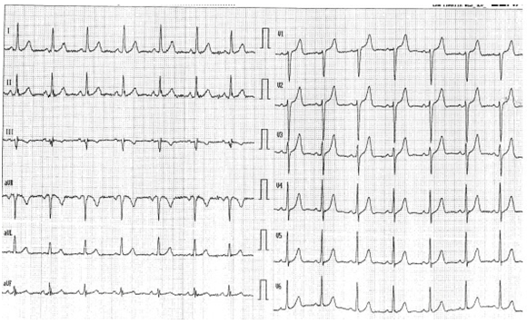
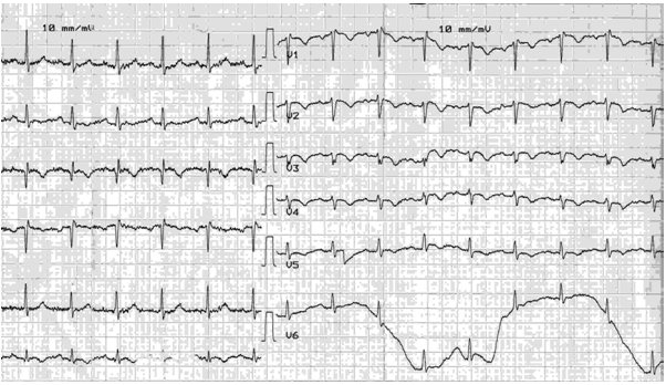
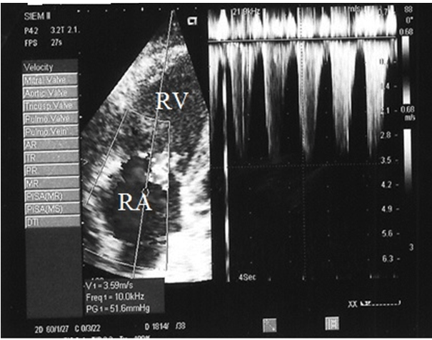
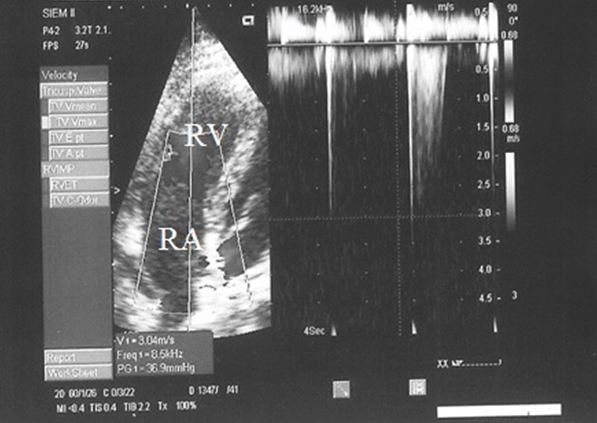
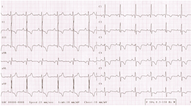
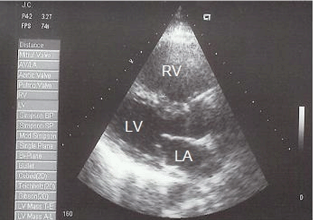
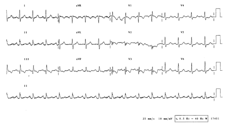
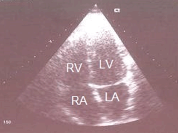
A 56-year-old white female presented without chronic disease, non-smoker, obese with body mass index 32% and with negative family history of venous thromboembolism (VTE). In 2002, she experienced acute pulmonary embolism (PE) treated by systemic thrombolysis (accelerated regimen of alteplase 100 mg over two hours) ( (Figure 1) (Figure 2) (Figure 3) (Figure 4), At this time, she used oral contraceptives for last five years, which were stopped and before discharge from hospital she received oral anticoagulation (vitamin K antagonists, warfarin). After six months of acute PE, she was examined for inherited thrombophilias associated with VTE and elevated levels of factor VIII 240 IU/dl (normal range 60–150 IU/dl) and homocystein 20.9 umol/l (normal range 5–12 umol/l) were revealed. The patient was recommended to continue with warfarin. One year after pulmonary embolism, exertional dyspnea occurred and the patient was repeatedly examined by pneumologist in 2005 because of its progression. She underwent fibre optics bronchoscopy along with bronchoalveolar lavage and lungs biopsy, because interstitial lung disease was suspected (according to result of high resolution computed tomography) these examinations excluded suspected diagnosis. She underwent a bone marrow aspirate and biopsy because of progressive polycythemia for last two years (before bone marrow aspirate and biopsy hemoglobin was 182 g/l (normal range 120–155 g/l), hematocrit 0.61 (normal range 0.36–0.47), according to results, polycythemia vera was excluded. Despite of progressive dyspnea and only boundary result from pulmonary function tests and negative chest radiograph the patient continued in treatment by a pneumologist as bronchitis chronica. In the beginning of 2006, the patient experienced sudden chest pain because of which, she was admitted to the hospital as a suspected acute coronary syndrome without ST segment elevation (Figure 5). Next day of hospitalization transthoracic echocardiography (TTE) was performed revealing pulmonary hypertension (PH) with systolic pulmonary artery pressure 105 mmHg and dilatation of right ventricle and atrium (Figure 6). At this time, level of D-dimer was 0.21 mg/L, which was in normal range and apart from high red blood cell count (hemoglobin 18.6 g/dl; normal range 12.0–15.5 g/dl), hematocrit 0.64; normal range 0.36–0.47, leucocytes 8.10x10⁹/l; normal range 3.90–10.00x10⁹/l; thrombocytes 376x109/l; normal range 140–400x109/l), other laboratory tests were within the normal values. Subsequently performed computed tomography pulmonary angiogram (CTPA) was negative. Despite negative CTPA, we supposed diagnosis CTEPH, because of which, she underwent ventilation-perfusion (V/Q) lung scintigraphy (Figure 7). At this time, patient was classified in the New York Heart Association (NYHA) functional class III. After patient consent, she was referred to the highly specialized centre for pulmonary artery hypertension. After additional diagnostic procedures were performed (pulmonary angiography and right heart catheterization) the diagnosis CTEPH was definitely confirmed and she underwent PEA. In the perioperative period, reduction of mean pulmonary artery pressure (57 versus 28 mmHg) was observed. Four months after PEA, patients was in the NYHA functional class I, ECG (Figure 8) shown regress of right ventricle hypertrophy, as well as TTE (Figure 9) shown regress of PH and regress of dilatation right ventricle and atrium. Laboratory investigations revealed normalisation of red blood cell count (hemoglobin 14.6 g/dl; normal range 120–15.5 g/dl, hematocrit 0.43; normal range 0.36–0.47, leucocytes 5.50x10⁹/l; normal range 3.90–10.00x10⁹/l), thrombocytes 223x109/l; normal value 140–400x109/l). Presently, the patient is classified in the NYHA functional class I and except warfarin she intakes no other drugs. | ||||||
| ||||||
| ||||||
| ||||||
| ||||||
| ||||||
| ||||||
| ||||||
| ||||||
| ||||||
|
Discussion
| ||||||
|
Chronic thromboembolic pulmonary hypertension (CTEPH) results from incomplete resolution of a pulmonary embolus predominantly major (central or proximal large) and is listed as distinct subgroup of pulmonary hypertension (group 4) [3] [4]. Chronic thromboembolic pulmonary hypertension is an underdiagnosed disorder, and the true prevalence is still unclear, the disorder has been demonstrated as late complication of patients who survive an acute PE with a cumulative incidence of 0.1–9.1% within two years of the event [4]. In clinical practice, it is important to differentiate acute PE from an acute episode superimposed on pre-existing CTEPH, because first clinical presentation of CTEPH may mimic acute PE [5]. A significant number of CTEPH cases may originate from asymptomatic VTE (4) or develop in the absence of previous acute PE [6]. The slowly progressive nature of the course of CTEPH allows right ventricle hypertrophy to ensue, which compensates for the increased pulmonary vascular resistance. However, because of progressive thrombosis or vascular changes in the "uninvolved" vascular bed, PH becomes progressive which leads to hypoxaemia [7] and chronic hypoxaemia leads to secondary polycythemia [8]. Patients with CTEPH typically present in either of two scenarios: patients may complain of progressive dyspnea on exertion, hemoptysis, and/or signs of right heart dysfunction including fatigue, palpitations, syncope, or edema after a single episode or recurrent episodes of overt PE. A "honeymoon period" between the acute event and the development of clinical signs of CTEPH is common and may last from a few months to many years [6]. The fibrin derived from patients with CTEPH seems resistant to lysis and D-dimer is not elevated and then to measure of D-dimer is not suitable for diagnosis and prognosis of CTEPH [9][10], then normal level of D-dimer should exclude CTEPH from acute PE. Chest radiography, pulmonary function tests, ECG and echocardiography are used in the initial assessment of suspected PH. In general, standard chest radiographs and pulmonary function tests have limited sensitivity and specificity and are insufficient to diagnose CTEPH [7]. Echocardiography at rest remains the best way to estimate elevated pulmonary pressures according to the level of systolic PAP estimated by the tricuspid regurgitant velocity and can reveal an enlarged right ventricle with abnormal contractility [11]. The V/Q lung scintigraphy remains the main first-line imaging modality for CTEPH, as it carries a 96–97% sensitivity and 90–95% specificity for the diagnosis and should be performed in all patients with PH to look for CTEPH, a negative result excludes the diagnosis with almost 100% certainty [12]. While multi-detector computed tomography (MDCT) is the investigation of choice for the diagnosis of acute PE, the investigation may be used as a complementary investigation but does not replace the V/Q lung scintigraphy or traditional pulmonary angiogram [13]. The CTPA alone cannot rule out CTEPH, but may help identify complications of the disease, such as pulmonary artery distension, resulting in left main coronary artery compression [14]. Pulmonary angiography remains a standard diagnostic tool in the assessment of patients with probable or definite CTEPH both to establish the diagnosis and to assess operability. Pulmonary angiography should be performed in conjunction with a diagnostic right heart catheterization, which is an essential diagnostic tool. The comparison of radiographic burden of disease with hemodynamics is a critical exercise in determining operability as well as surgical risk before PEA. Both right heart catheterization and pulmonary angiography should be performed by experienced staff [15]. All patients with established CTEPH should receive life-long oral anticoagulants, unless contraindicated, while no data exist on the efficacy and safety of new direct oral anticoagulants. PEA is the only effective treatment for eliminating the cause of the disease [3]. The effective PEA is associated with the reduced mortality and leads to a permanent improvement in the pulmonary hemodynamics and exercise capacity of patients. In Europe, in-hospital mortality is currently as low as 4.7% in expert centers [16]. Therefore, all patients with CTEPH should be referred for operability assessment by an experienced CTEPH team to determine if the patient is operable and candidate for PEA. If a patient is deemed non-operable, this patient should be repeatedly referred for operability assessment for a second opinion by an experienced CTEPH team [17] [18]. The highly specialized centre for pulmonary artery hypertension for Slovakia is Cardio Centre of General University Hospital in Prague [19]. For patients deemed non-operable by PEA other treatment options in select cases may include lung transplantation or percutaneous transluminal pulmonary angioplasty [16] [17]. Pharmacological therapy should be considered in patients with inoperable/persistent CTEPH after PEA, who face a poor prognosis. Currently, riociguat (soluble guanylate cyclase stimulator) is the only registered drug [20][21][22]. | ||||||
|
Conclusion
| ||||||
|
Chronic thromboembolic pulmonary hypertension (CTEPH) is a rare complication of acute pulmonary embolims (PE), but it can also occur in patients who do not have a history of acute PE or deep vein thrombosis (DVT) is under-diagnosed and also frequently misdiagnosed in clinical practice. The present report aims to increase the awareness of clinicians towards an accurate diagnosis of the disease. Ventilation-perfusion (V/Q) lung scintigraphy should be the basic and the first diagnostic tool. A negative result virtually excludes the diagnosis with almost 100% certainty. At the same time, the presence of perfusion defects in scintigraphy does not confirm CTEPH. Further diagnostics is necessary, which should involve a number of studies, including right heart catheterization and pulmonary angiography. Each patient diagnosed with CTEPH should be considered for PEA, as it is the only effective treatment method for eliminating the cause of the disease, leading to cure. | ||||||
|
References
| ||||||
| ||||||


|
[HTML Abstract]
[PDF Full Text]
|
|
Author Contributions
Margita Belicová – Substantial contributions to conception and design, Acquisition of data, Analysis and interpretation of data, Drafting the article, Revising it critically for important intellectual content, Final approval of the version to be published Veronika Jankovičová – Analysis and interpretation of data, Revising it critically for important intellectual content, Final approval of the version to be published Marian Mokáň – Analysis and interpretation of data, Revising it critically for important intellectual content, Final approval of the version to be published |
|
Guarantor of submission
The corresponding author is the guarantor of submission. |
|
Source of support
None |
|
Conflict of interest
Authors declare no conflict of interest. |
|
Copyright
© 2017 Margita Belicová et al. This article is distributed under the terms of Creative Commons Attribution License which permits unrestricted use, distribution and reproduction in any medium provided the original author(s) and original publisher are properly credited. Please see the copyright policy on the journal website for more information. |
|
|