| |
|
|
|
Case Report
| ||||||
| The case of an extensive primary extramammary Paget's disease diagnosis and treatment | ||||||
| Apostolos Sarivalasis1, Cécile Triboulet2, Sandro Anchisi3 | ||||||
|
1Consultant, Department of medical oncology, Centre Hospitalier du Valais Romand (CHVR), Sion, Switzerland.
2Dermato-Pathologist. Department Dermatology and dermatopathology, Centre Hospitalier Universitaire Vaudois (CHUV), Lausanne, Switzerland. 3Head Oncologist, Director. Department of medical oncology, Centre Hospitalier du Valais Romand (CHVR), Sion, Switzerland. | ||||||
| ||||||
|
[HTML Abstract]
[PDF Full Text]
[Print This Article]
[Similar article in Pumed] [Similar article in Google Scholar] 
|
| How to cite this article |
| Sarivalasis A, Triboulet C, Anchisi S. The case of an extensive primary extramammary Paget's disease diagnosis and treatment. Int J Case Rep Images 2017;8(1):46–50. |
|
Abstract
|
|
Introduction:
Extramammary Paget's disease (EMPD) is a rare neoplasm commonly related with underlying malignancy. It mainly affects intimate body areas and presents with eczema like lesions. Due to the rarity, the indolent natural history and the anatomical distribution of this disease, the diagnosis is difficult and often delayed. Treatment can be challenging especially since the majority of patient are elderly and frail.
Case Report: A 91-year-old male suffering from coronary disease and myelodysplastic syndrome presented with a slowly evolving scrotal eruption. A differential diagnosis was established and a punch biopsy was performed. An EMPD was diagnosed. The known relation of EMPDs with underlying malignancies mandated further complementary examinations. An underlying malignancy was excluded and a personalized treatment was undertaken. Conclusion: Extramammary Paget's disease diagnosis and treatment are challenging. A comprehensive differential diagnosis is essential for integrating this rarity in the diagnostic assessment of persistent eczematous eruptions. A skin biopsy is paramount for diagnosis. A guided workout with multimodal examinations to rule out underlying malignancies is strongly recommended. The gold standard of EMPD treatment is surgery. Nevertheless, alternative less invasive treatments should be considered, depending on patient's comorbidities. | |
|
Keywords:
Elderly, Extramammary Paget's disease, Skin rash
| |
|
Introduction
| ||||||
|
Cutaneous Paget's disease is a rare intraepithelial neoplastic condition [1] [2]. The breast Paget's disease can be associated with invasive and in situ breast cancer and is a well-defined entity. The same histological pattern (Paget's cells) [3] can be found in other body areas with abundance of apocrine glands such as the perineum, scrotum, vulva, penis, and axilla [4]. In such cases it is called extramammary Paget's disease (EMPD). The EMPD affects mostly elderly persons and can be primary or secondary. Primary EMPD originate from intraepidermal cells. Secondary EMPD is associated with underlying malignancy and accounts for 10–30% of all EMPD cases series [1] [5][6]. The most common associated neoplasms arise from the bladder, urethra, prostate and rectum. The involved body area can help guide diagnostic procedures since the bladder, urethra, and prostate cancers are associated with EMPDs of the external genitalia, while rectal adenocarcinoma is commonly related with perianal EMPD [6]. The relatively indolent natural history, the lack of specific symptoms and the body distribution (most frequently in the anogenital region followed by the axilla) often delays diagnosis, resulting in extensive lesions. The treatment of those lesions can be challenging. Different procedures have been reported effective with surgery being considered the standard of care [5] [7] [8] . This radical approach cannot be considered in every clinical setting. Hence, alternative local treatments have been evaluated. Imiquimod 5% topical cream [9][10][11], photodynamic therapy [12] [13]and radiation therapy [14] are the most frequently applied alternative treatments. | ||||||
|
Case Report
| ||||||
|
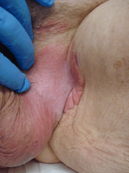
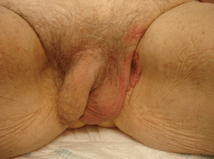
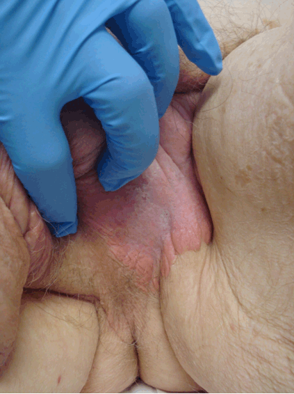
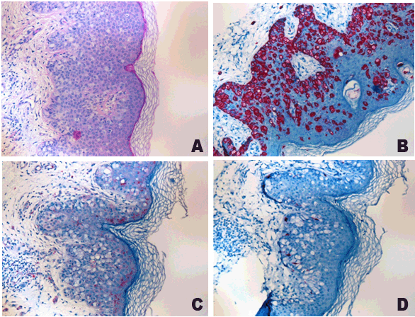
A 91-year-old male known for refractory cytopenia myelodysplastic syndrome and coronary disease, presented with a slowly evolving scrotal and perineal eruption. The onset of the eruption could not be dated. Previously prescribed corticosteroids and hydrating local treatments were ineffective. The lesion covered approximately 1% of the body surface and infiltrated the scrotum, the left lateral perineal area extending to 2 cm from the anal margin (Figure 1) (Figure 2) (Figure 3). Two smaller satellite infiltrated areas were noted in the left lower inguinal region (Figure 4). The affected skin area was evenly infiltrated and erythematous. Areas of desquamation were present and the eruption borders presented swelling. The lesion was neither tender nor painful and the patient's main complaint was intermittent pruritus. Neither lymphadenopathy nor hepatosplenomegaly was noted. The digital rectal examination and external genitalia inspection were normal. The histological examination of a punch biopsy specimen showed a typical image of Paget's cells in hematoxylin and eosin coloration (Figure 5A). In immunohistochemistry (IHC) the cells were cytokeratine 7 (CK7) positive (Figure 5B) while S100 (Figure 5C) and Melan A were both negative (Figure 5D). After the histological confirmation, a comprehensive diagnostic assessment to rule out underlying malignancies was ordered. The whole body CT scan and the urological examination were free of pathological findings. The gold standard of EMPD treatment being a wide surgical resection a surgical assessment was conducted. Due to the disease extension, the patient's age and the concurrent medical conditions an upfront surgical treatment was deemed not appropriate. Radiotherapy was also ruled out according to the patient's desire. A topical approach with either photodynamic therapy or local immunomodulatory cream application, considered as less invasive, was presented to the patient [12][13][15][16]. None of these treatments are as effective as surgery but promising results are reported in literature, although controversial. The benefit-risk ratio of these approaches being judged favorably by the patient he was addressed to the Lausanne's University Dermatological Hospital. Because of the extent of the lesion, too large to be treated with local immunomodulator application of imiquimod cream 5%, a photodynamic therapy was prescribed. Six months disease's stabilization was observed on this treatment. | ||||||
| ||||||
| ||||||
| ||||||
| ||||||
| ||||||
|
Discussion
| ||||||
|
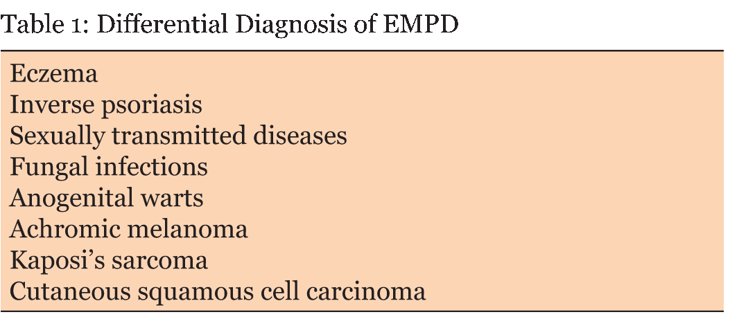
In this case, the differential diagnosis included several skin conditions (Table 1). The pruritus, as the leading symptom, can be misdiagnosed for eczema. However the extent and characteristics of the eruption, the absence of relevant history or clinical findings of atopia and the lack of response to front line eczema treatment suggested an alternative diagnosis. Inverse psoriasis needs also to be considered as differential diagnosis since it affects intertriginous areas and often appears without the typical scaly lesions. Fungal infections can grow involving the same anatomical regions as EMPD. Fungal lesions are small sized, but when confluent can merge to larger areas. A skin sample usually provides the diagnosis. Sexually transmitted diseases (STDs) such as Syphilis needs to be ruled out by screening tests and other serological and bacteriological examinations. Anogenital warts are often small and asymptomatic but can, if neglected, grow into large exophytic conglomerates appearing as papilliform masses. A nonspecific test with 5% acetic acid can be useful for the differential diagnosis highlighting wart lesions in white. This was not the patient's case. Achromic melanoma is a rarity. It presents as a non-pigmented skin lesion and often has a more aggressive natural history with early metastasis, notably, to lymph nodes. Kaposi's sarcoma involves mostly extremities, exhibits an aggressive behavior and presents with characteristic purple lesions. Nevertheless, due to morphologic variants a biopsy should always be performed. Another dermato-oncological diagnosis to be ruled out is cutaneous squamous cell carcinoma, an entity that diagnosis exhibits distinct pathological features. To exclude alternative neoplastic skin disorder a pathological assessment is paramount. The EMPD growth characteristics, including large epithelial cells proliferation- Paget's cells - distributed in small clusters between normal keratinocytes and typical IHC expression is the cornerstone for diagnosis. The known association of EMPD with underling malignancies mandates a thorough assessment with appropriate laboratory, endoscopic and radiological tests. Our patient suffered from a primary EMPD since no underlying malignancy was diagnosed on the diagnostic assessment. The treatment approach was personalized taking into account the medical history, age, expected morbidity and patient's choice. The photodynamic treatment was proved to be an effective therapeutic option with very good local results. | ||||||
| ||||||
|
| ||||||
|
Conclusion
| ||||||
|
Extramammary Paget's disease (EMPD) is a rare neoplasm. The initial presentation can be misleading and easily misdiagnosed. High level of clinical awareness and histological confirmation are essentials in order to obtain correct diagnosis. A comprehensive diagnostic assessment is highly recommended because of the known association of EMPD with underling malignancies. The treatment plan needs to be personalized especially in elderly and frail patients where concomitant health conditions contraindicate extensive surgical management of the disease. | ||||||
|
References
| ||||||
| ||||||
|
[HTML Abstract]
[PDF Full Text]
|
|
Author Contributions
Apostolos Sarivalasis – Substantial contributions to conception and design, Acquisition of data, Analysis and interpretation of data, Drafting the article, Revising it critically for important intellectual content, Final approval of the version to be published Cécile Triboulet – Substantial contributions to conception and design, Acquisition of data, Analysis and interpretation of data, Final approval of the version to be published Sandro Anchisi – Substantial contributions to conception and design, Drafting the article, Revising it critically for important intellectual content, Final approval of the version to be published |
|
Guarantor of submission
The corresponding author is the guarantor of submission. |
|
Source of support
None |
|
Conflict of interest
Authors declare no conflict of interest. |
|
Copyright
© 2017 Apostolos Sarivalasis et al. This article is distributed under the terms of Creative Commons Attribution License which permits unrestricted use, distribution and reproduction in any medium provided the original author(s) and original publisher are properly credited. Please see the copyright policy on the journal website for more information. |
|
|