| |
|
|
|
Case Report
| ||||||
| Intraosseous acinic cell carcinoma: A rare case report | ||||||
| Lakshmana N.1, Vamsi Pavani B.2, Abhishek Singh Nayyar3, Kartheeki B.4, Kalyana Chakravarthy B.5, Kameswara Rao A.6 | ||||||
|
1Senior Lecturer, Department of Oral Medicine and Radiology, Sri Sai Dental College and Hospital, Srikakulam, Andhra Pradesh, India.
2Professor and Head, Department of Oral Medicine and Radiology, Sri Sai Dental College and Hospital, Srikakulam, Andhra Pradesh, India. 3Reader cum PG Guide, Department of Oral Medicine and Radiology, Saraswati Dhanwantari Dental College and Hospital and Post-Graduate Research Institute, Parbhani, Maharashtra, India. 4Senior Lecturer, Department of Oral Medicine and Radiology, Saraswati Dhanwantari Dental College and Hospital and Post-Graduate Research Institute, Parbhani, Maharashtra, India. 5Reader, Department of Oral Medicine and Radiology, Sri Sai Dental College and Hospital, Srikakulam, Andhra Pradesh, India. 6Assistant Dental Surgeon, Community Health Center, Vizianagaram, Andhra Pradesh, India. | ||||||
| ||||||
|
[HTML Abstract]
[PDF Full Text]
[Print This Article]
[Similar article in Pumed] [Similar article in Google Scholar] 
|
| How to cite this article |
| Lakshmana N, Vamsi PB, Nayar AS, Kartheeki B, Chakravarthy KB, Rao KA. Intraosseous acinic cell carcinoma: A rare case report. Int J Case Rep Images 2017;8(1):36–40. |
|
Abstract
|
|
Introduction:
De-differentiated acinic cell carcinoma of salivary glands is an uncommon variant of acinic cell carcinoma characterized by the co-existence of both low grade acinic cell carcinoma and a high-grade de-differentiated component as well as an aggressive clinical course.
Case Report: Herewith, we are reporting a case of de-differentiated acinic cell carcinoma which was present in mandibular region. A 35-year-old female patient reported with a chief compliant of a swelling since one month and pain since 15 days in the lower left back tooth region. To the best of our knowledge, this location has never been described in de-differentiated acinic cell carcinoma as parotid is the most common site for its occurrence. Conclusion: Despite the rarity of intraosseous acinic cell carcinomas, one should be well aware of this diagnostic possibility, emphasizing the need for histopathological analysis, a clearly defined and guided treatment strategy, and an adequate follow-up to check for the possibility of any recurrences. | |
|
Keywords:
De-differentiation, Low grade acinic cell carcinoma
| |
|
Introduction
| ||||||
|
Acinic cell carcinoma is a neoplasm of low grade malignancy and composed of cells that have got differentiated towards serous acinar cells. It was originally described by Nasse in 1892 as a low grade, benign lesion with later studies confirming its malignant behavior which was in between that of adenoma and carcinoma [1]. Hence, it was initially termed acinic cell tumor in the earlier WHO classification in 1972 which was later revised to acinic cell carcinoma in 1991. Acinic cell carcinoma is considered to be the third most common major salivary gland tumor/malignancy [2]. De-differentiation or, high-grade malignant transformation (HGT) has been described in a variety of salivary gland tumors although the phenomenon is reported to be a relatively rare event. Authors preferably use the term HGT rather than de-differentiation for such cases [3]. De-differentiation is the progression of cells towards a less differentiated state in which the original line of differentiation is no longer evident. The first acinic cell carcinoma with high grade malignant transformation of salivary gland was reported by Stanley et al. in 1988. Thirty-five cases have been described in literature so far and most of them showed poor clinical outcome. Also, all cases reported to date were of parotid gland origin with involvement of both the superficial and/or, deep lobes. These tumors have a slight male predisposition, high recurrence rates, and a high propensity for cervical lymph node metastasis, suggesting a role for neck dissection in the management of affected patients [4] . Furthermore, vascular and peri-neural invasions are typically observed in acinic cell carcinomas. The diagnosis is usually confirmed with a fine needle aspiration cytology (FNAC) procedure, while radical surgical excision of the tumor is the mainstay of treatment of this malignant neoplasm. Other treatment modalities include radiotherapy which might be indicated in some cases. Acinic cell carcinomas have a significant tendency to recur, to lead to metastases in cervical lymph nodes, and lungs rarely, and may have an aggressive evolution, therefore, making long-term follow-up, mandatory, post-treatment. | ||||||
|
Case Report
| ||||||
|
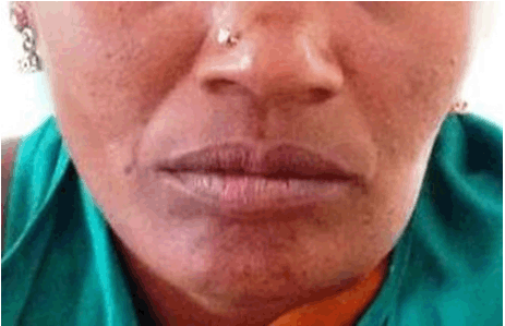
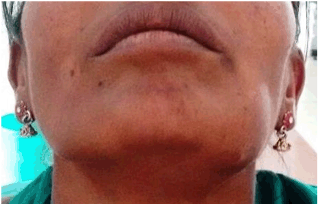
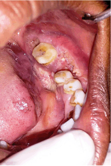
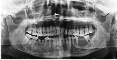
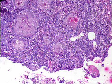
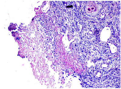
A 35-year-old female reported with a chief compliant of a swelling since one month and pain since 15 days in the lower left back tooth region. The swelling actually had an insidious origin and progression and started as a small imperceptible growth which was painless initially and gradually increased in size and became painful with the pain being continuous, dull, throbbing in nature which used to get aggravated on brushing and other mechanical trauma/provocation and on taking hard foods. Pain used to get temporarily relieved with medication. There was shedding of a tooth in the left lower back tooth region 15 days prior to the reporting of the patient. Since then, patient gave a history of increase in the size of the swelling. Patient had multiple, palpable submental and submandibular lymph nodes, present which were firm, mobile and tender. On clinical examination, there was a single, unilateral, ovoid-shaped, swelling present irt left lower third of face measuring approximately 4x5 cm in greatest dimensions (Figure 1). The skin overlying the swelling was normal in appearance without signs of any erythema and/or, discharge or, ulceration. The swelling had well-defined edges and the borders extended from 2 cm away from corner of the mouth anteriorly to 2 cm ahead of the angle of mandible posteriorly, and from below the imaginary line drawn from the corner of the mouth and lobule of ear superiorly to approximately 1 cm below the inferior border of the mandible inferiorly (Figure 2). On palpation, the swelling was well-defined, firm to hard in consistency, slightly tender and fixed to the underlying bone. On intra-oral examination, there was an ulcero-proliferative, exophytic growth seen irt teeth #35, #36, #37 region on the lingual side. The mucosa was perforated due to expansion of the buccal and lingual cortical plates with exposure of the white, necrosed bone (Figure 3). On palpation, tenderness and vestibular obliteration were present. Orthopantomograph (OPG) revealed a solitary, irregular radiolucency extending from the distal surface of tooth #34 till the mesial surface of tooth #37. (Figure 4). Incisional biopsy was performed and sent for histopathological examination which showed neoplastic cells arranged in solid, lobular pattern, separated by thin, fibrous connective tissue septae (Figure 5). Epithelial cells were pleomorphic in nature with increased mitotic activity and with keratin pearl formation suggestive of a de-differentiated acinic cell carcinoma (Figure 6). Based on the said clinical, radiological and histopathological features, a final diagnosis of a primary intraosseous salivary gland carcinoma was arrived-at. The patient was, then, referred for hemimandibulectomy and re-construction followed by radiotherapy and chemotherapy under guidance. | ||||||
| ||||||
|
| ||||||
| ||||||
| ||||||
| ||||||
| ||||||
|
Discussion
| ||||||
|
Acinic cell carcinoma is a low grade malignant epithelial neoplasm of salivary gland tissue origin in which at least few of the neoplastic cells demonstrate serous acinar differentiation characterized by the presence of cytoplasmic zymogen secretory granules. These carcinomas account for about 4% of all salivary gland neoplasms with around 7–17.5% going for malignant transformation [5]. Numerous reports indicate primary salivary gland neoplasms to be completely intra-bony, yet, non-neoplastic salivary gland tissues have rarely been found in such locales, with few reports suggesting odontogenic origin of such tumors, although, the rate of occurrence of salivary gland choristomas, hamartomas, embryonic rests, and aberrant salivary gland tissues within the alveolar bone, is less than 2.6 of every 1000 marrow samples biopsied with this providing an additional and quite seemingly logical histogenetic explanation for the presence of intraosseous salivary gland neoplasms [6]. Neoplastic transformation of intra-bony salivary gland tissues, in particular, is an uncommon phenomenon with mandible being one of the most common sites for such neoplasia, accounting for around 75% of all the cases seen [7]. Entrapment of salivary gland tissue within the jaw bones during initial stages of development and metaplastic transformation of the epithelial lining of odontogenic cysts are the two significant hypotheses that have been proposed for such rare, intraosseous salivary gland neoplasms [8]. Acinic cell carcinomas may be found in all age groups, including children and early adolescence, with the peak incidence noted in the fifth and sixth decades of life. The most important clinical features include slow-growing lesions with an insidious onset and progression, most of the times, and which are attended by pain, later, in the course of development due to secondary changes and/or, as a result of mechanical trauma after they reach sufficient sizes [9]. Histopathologically, large lobules or, nests of tumor cells with little intervening stroma are characteristic. The arrangement of neoplastic cells is quite variable. Generally, cells are arranged in solid masses with blunted or, pushing margins. Other variants include microcystic, papillary, and follicular forms. Radical surgical excision of the tumors is the preferred treatment although recurrences are not uncommon [10]. | ||||||
|
Conclusion
| ||||||
|
To conclude, despite the rarity of intraosseous acinic cell carcinomas, one should be well aware of this diagnostic possibility, emphasizing the need for histopathological analysis, a clearly defined and guided treatment strategy, and an adequate follow-up to check for the possibility of any recurrences. | ||||||
|
References
| ||||||
| ||||||
|
[HTML Abstract]
[PDF Full Text]
|
|
Author Contributions
Lakshmana N. – Substantial contributions to conception and design, Acquisition of data, Analysis and interpretation of data, Drafting the article, Revising it critically for important intellectual content, Final approval of the version to be published Vamsi Pavani B. – Analysis and interpretation of data, Revising it critically for important intellectual content, Final approval of the version to be published Abhishek Singh Nayyar – Analysis and interpretation of data, Revising it critically for important intellectual content, Final approval of the version to be published Kartheeki B. – Analysis and interpretation of data, Revising it critically for important intellectual content, Final approval of the version to be published Kalyana Chakravarthy B. – Analysis and interpretation of data, Revising it critically for important intellectual content, Final approval of the version to be published Kameswara Rao A. – Analysis and interpretation of data, Revising it critically for important intellectual content, Final approval of the version to be published |
|
Guarantor of submission
The corresponding author is the guarantor of submission. |
|
Source of support
None |
|
Conflict of interest
Authors declare no conflict of interest. |
|
Copyright
© 2017 Lakshmana N. et al. This article is distributed under the terms of Creative Commons Attribution License which permits unrestricted use, distribution and reproduction in any medium provided the original author(s) and original publisher are properly credited. Please see the copyright policy on the journal website for more information. |
|
|