
  |

|
 |
|
Case Report
| ||||||
| Rare presentation of a massive intermittent upper gastrointestinal bleed | ||||||
| Bonnie Patek1, Matthew Sullivan1, Shashin Shah2 | ||||||
|
1DO, Internal Medicine Resident, Department of Internal Medicine, Lehigh Valley Health Network, Allentown, PA, USA.
2MD, Department of Gastroenterology, Lehigh Valley Health Network, Allentown, PA, USA. | ||||||
| ||||||
|
[HTML Abstract]
[PDF Full Text]
[Print This Article]
[Similar article in Pumed] [Similar article in Google Scholar] 
|


| How to cite this article |
| Patek B, Sullivan M, Shah S. Rare presentation of a massive intermittent upper gastrointestinal bleed. Int J Case Rep Images 2017;8(1):30–35. |
|
Abstract
|
|
Introduction:
Hemosuccus pancreaticus is a rare cause of upper gastrointestinal bleeding defined as bleeding from the ampulla of Vater through the main pancreatic duct, commonly caused by a ruptured aneurysm in the setting of acute or chronic pancreatitis. Bleeding is often intermittent and repetitive, but can be massive. Endoscopy rarely reveals active hemorrhaging.
Case Report: We present a 35-year-old male with complaints of progressive fatigue and generalized weakness over two weeks. He denied melena, hematochezia, and hematemesis. History revealed prior alcohol abuse and hospitalization for pancreatitis. Laboratory studies revealed hemoglobin of 2.8 g/dL. Initial esophagogastroduodenoscopy (EGD) revealed actively bleeding mass at the major papilla and absence of gastric/esophageal varices. Colonoscopy revealed no pathology. Bleeding continued and patient developed mild epigastric tenderness. Repeat EGD revealed a clean based ulcer in the distal esophagus and thickening of the duodenal mucosa, but no bleeding. Duodenal biopsies showed signs of chronic inflammation. Abdominal computed tomography scan revealed pancreatitis and splenic artery pseudoaneurysm. Patient underwent an abdominal aortogram, revealing an 8 mm splenic artery pseudoaneurysm with no active bleeding. The pseudoaneurysm was embolized and repeat imaging confirmed no residual pseudoaneurysm. Conclusion: Pseudoaneurysm resulting from a pseudocyst secondary to pancreatitis forms most commonly in the splenic artery. Mortality can be as high as 90–100% if left untreated. Although rare, hemosuccus pancreaticus should be included in the differential diagnosis for any patient presenting with severe anemia, in the absence of endoscopically visualized bleeding and with a history of pancreatitis. | |
|
Keywords:
Hemosuccus pancreaticus, Pancreatitis, Splenic artery pseudoaneurysm, Upper gastrointestinal bleeding
| |
|
Introduction
| ||||||
|
Hemosuccus pancreaticus is a rare cause of upper gastrointestinal bleeding defined as bleeding from the ampulla of Vater through the main pancreatic duct, commonly caused by a ruptured aneurysm in the setting of acute or chronic pancreatitis. This terminology was described in 1970 by Dr. Sandblom and characterized by gastrointestinal bleeding leading to the cause of anemia and possible sharp episodic epigastric pain that can easily be overlooked, originating from a splenic artery aneurysm that ruptured into the main pancreatic duct [1]. Aneurysms and pseudoaneurysms are commonly formed in the setting of both acute and chronic pancreatitis [2]. However, these are not the only causes of hemosuccus pancreaticus (Table 1) [2][3][4][5][6][7]. Bleeding in these cases is often intermittent and repetitive, but can be massive. The intermittent bleeding of hemosuccus pancreaticus, despite being from an arterial source, rarely presents with hemodynamic instability, unless the cause is from a pseudoaneurysm or aneurysm that has ruptured [2]. The intermittent bleeding makes the diagnosis difficult with the possible need for multiple imaging modalities and high index of clinical suspicion to continue to look for a bleeding source. Endoscopy rarely reveals active hemorrhaging and often CT scan or angiography is needed to locate the source of the hemorrhage. If the source is located, interventional radiology should be consulted and proceed with embolization [5]. If these are unsuccessful or the patient is hemodynamic unstable, surgical intervention with the use of intraoperative pancreatoscopy is necessary to determine and treat the hemorrhagic source [6]. | ||||||
| ||||||
|
| ||||||
|
Case Report
| ||||||
|
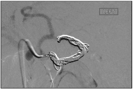
A 35-year-old male presented with a past medical history pertinent only for an occult gastrointestinal bleed one year ago during a bout of pancreatitis. Patient complained of generalized weakness and progressive fatigue for the last two weeks. He denied melena, hematochezia, hematemesis, nausea/vomiting, jaundice or chronic ibuprofen usage. Further history revealed prior alcohol abuse discontinued two years ago and a hospitalization for acute pancreatitis about four years ago. Laboratory studies revealed hemoglobin of 2.8 g/dL, which was followed by a blood transfusion of four packed red blood cell (pRBC) units. An actively bleeding mass was seen at the major papilla during the initial esophagogastroduodenoscopy (EGD). No other sources of bleeding were noted as the patient had a non-bleeding ulceration at the gastroesophageal junction and lacked gastric or esophageal varices. A colonoscopy was performed that was unremarkable for a bleeding source. These procedures were performed prior to transfer to our institution. Bleeding continued with a continued drop in hemoglobin. Upon arrival to our institution, the patient was noted to have spiking fevers and developed acute epigastric tenderness. Repeat hemoglobin was 7.1 g/dl leading to another two units of pRBC being transfused. An elevated lipase level at 803 U/L was noted. A repeat EGD was performed at our institution and revealed no mass at the major papilla or source of bleeding. Mild thickening of the duodenal sweep and a clean based ulcer at the distal esophagus were the only findings. Duodenal biopsies exhibited only signs of chronic inflammation. A computed tomography scan of the abdomen/pelvis revealed pancreatitis consistent with splenic artery pseudoaneurysm (Figure 1), splenomegaly and 10x12 mm pancreatic head lesion (Figure 2) that was non-specific. The patient was evaluated by interventional radiology and underwent an abdominal angiography, revealing an 8 mm splenic artery pseudoaneurysm from the proximal-mid splenic artery with no active bleeding. Coil embolization with six clips (Figure 3) and repeat imaging confirmed no residual pseudoaneurysm (Figure 4). After the procedure, the patient experienced mild abdominal discomfort, fevers, leukocytosis which could be secondary to a splenic infarction (Figure 5) that was also observed on CT scan status post embolization or from resolving pancreatitis. Follow-up MRI scan to evaluate the pancreatic mass showed an ill-defined area of decreased attenuation measuring 1.3 cm in addition to fatty infiltrates within the liver and splenic infarct. Patient was advised to follow- up for endoscopic ultrasound to evaluate this lesion after his acute issue had resolved. At time of discharge, patient was afebrile with no abdominal tenderness, stable hemoglobin and was vaccinated for encapsulated bacteria secondary to his asplenic condition status post splenic infarct. | ||||||
| ||||||
| ||||||
| ||||||
| ||||||
|
| ||||||
|
Discussion
| ||||||
|
Hemosuccus pancreaticus is a rare cause of upper gastrointestinal bleeding. There is a male predominance (7:1) associated with this disease. Pathophysiology is either from direct rupture of the aneurysm or pseudoaneurysm (PA) into the main pancreatic duct or indirect communication between the artery and duct with the pseudocyst. Weakening of the vessel wall forms the PA and is due to constant pressure necrosis (pseudocyst) and autodigestion from leakage of pancreatic enzymes from the pancreatitis [2] . Pseudoaneurysms commonly form in the setting of chronic (most common) or acute pancreatitis. Pseudoaneurysms form most commonly in the splenic artery (60%) followed by gastroduodenal (20%), pancreaticoduodenal (10%), hepatic (5%) and left gastric (2%) arteries in descending frequency. Complications of aneurysms and PAs include rupture leading to hemorrhage into the gastrointestinal tract, a pseudocyst, peritoneal cavity, retroperitoneal space or adjacent organs. The risk of rupture for an aneurysm is dependent on size, but there is no correlation in size with rupture of PAs or predictability therefore, all PAs should be treated. Frequency of rupture in the setting of pancreatitis is 5–10% and with a pseudocyst present, this can rise up to 20%. Mortality of a ruptured PA can be as high as 90–100% and if treated, the mortality risk is reduced to 12–57% [7] [8][9][10] . Bleeding is often intermittent, possibly due to clotting occurring within the main pancreatic duct, consequentially making diagnosis with endoscopy difficult [9]. Esophagogastroduodenoscopy is the first modality in order to rule out other more common causes of upper gastrointestinal bleeding. If bleeding is observed from the major ampulla, this is a strong indicator of hemosuccus pancreaticus, though seldom seen. Other signs may include residual blood clots located near the ampulla, which may be difficult to visualize with no signs of peptic ulcer disease, varices or gastritis [7]. If no clear bleeding source, CT scan of the abdomen is the first line imaging modality of choice to identify pancreatic pathology followed-by angiography, which is considered the gold standard for detecting PAs and first line treatment [5][10] . Embolization is first line therapy in hemodynamically stable patients. Coil embolization has a 79–100% success rate and a mortality rate of 12–33%. Both coils and glue are acceptable, coiling having less success if the vessel has increased tortuosity. Complications include embolization of undesired vessel, ischemia of organs with a lack of collateral circulation, infection and splenic infarct in the specific case of splenic artery involvement. Some of these complications can be avoided with the use of a non-coated metallic stent and the stent can be used as bridge to surgery in high risk patients or those with pancreatic disease that is in need of surgical treatment. The advantages to embolization include precise location, assessment of proper collateral flow, and excellent alternative for patients that are poor surgical candidates [7][8][9][10] . If hemodynamically unstable or embolization has failed or re-bleeding has occurred, surgery is recommended. Surgery depends on the location of the bleeding but most common procedures include bipolar arterial ligation, direct intra-pseudocystic ligation with pseudocyst drainage [9]. Pancreatic resection maybe necessary to control the pancreatic disease and the arterial bleeding with such operations as pancreaticoduodenectomy or splenopancreatectomy preferred in the setting of chronic pancreatitis [5]. The incidence of hemosuccus pancreaticus may rise in the future as a result of increasing alcohol use, primarily in males in the western countries consequentially causing more cases of pancreatitis, which could possibly, in turn, cause more occurrence of PAs leading to cases of hemosuccus pancreaticus [11]. Hemosuccus pancreaticus should rank higher on a differential diagnosis in the presence of chronic or even acute pancreatitis especially in the setting of chronic alcohol use in males. Given that the most common etiology for pancreatitis in the United States is alcohol induced and with the incidence continuing to rise, the increase in laparoscopic surgeries and the use of EUS-FNA for suspicious lesions residing in the pancreas, the incidence of hemosuccus pancreaticus may increase in the future. | ||||||
|
Conclusion
| ||||||
|
Hemosuccus pancreaticus is a rare cause of an upper gastrointestinal bleed that is difficult to diagnose with endoscopy alone and often requires multiple imaging modalities to visualize. It should be included in a differential for any patient that presents with melena, generalized weakness and fatigue with low hemoglobin that cannot be explained by more common causes such as peptic ulcer disease, varices, gastritis or iron deficiency anemia with colon cancer ruled out if the patient is over the age of 50. Treatment is based on the hemodynamic stability of the patient and the ability to identify the bleeding source, but embolization should be attempted prior to surgical interventions. | ||||||
|
Acknowledgements
| ||||||
|
We would like to acknowledge the internal medicine and gastroenterology departments at Lehigh Valley Health Network for support in submitting and presenting this case report. | ||||||
|
References
| ||||||
| ||||||




|
[HTML Abstract]
[PDF Full Text]
|
|
Author Contributions
Bonnie Patek – Substantial contributions to conception and design, Acquisition of data, Analysis and interpretation of data, Drafting the article, Revising it critically for important intellectual content, Final approval of the version to be published Matthew Sullivan – Analysis and interpretation of data, Revising it critically for important intellectual content, Final approval of the version to be published Shashin Shah – Analysis and interpretation of data, Revising it critically for important intellectual content, Final approval of the version to be published |
|
Guarantor of submission
The corresponding author is the guarantor of submission. |
|
Source of support
None |
|
Conflict of interest
Authors declare no conflict of interest. |
|
Copyright
© 2017 Bonnie Patek et al. This article is distributed under the terms of Creative Commons Attribution License which permits unrestricted use, distribution and reproduction in any medium provided the original author(s) and original publisher are properly credited. Please see the copyright policy on the journal website for more information. |
|
|
|
About The Authors
| |||
| |||
| |||
| |||
