
  |

|
 |
|
Clinical Image
| ||||||
| Inferior vena cava agenesis with exuberant collateral circulation | ||||||
| Ana Vaz Cristino1, Renata Silva1, Carmen Pais1, José Presa2 | ||||||
|
1MD, Resident, Internal Medicine, Centro Hospitalar de Trás-os-Montes e Alto Douro, Vila Real.
2MD, Attending, Internal Medicine, Centro Hospitalar de Trás-os-Montes e Alto Douro, Vila Real. | ||||||
| ||||||
|
[HTML Abstract]
[PDF Full Text]
[Print This Article]
[Similar article in Pumed] [Similar article in Google Scholar] 
|


| How to cite this article |
| Cristino AV, Silva R, Pais C, Presa J. Inferior vena cava agenesis with exuberant collateral circulation. Int J Case Rep Images 2017;8(1):86–88. |
|
Case Report
| ||||||
|
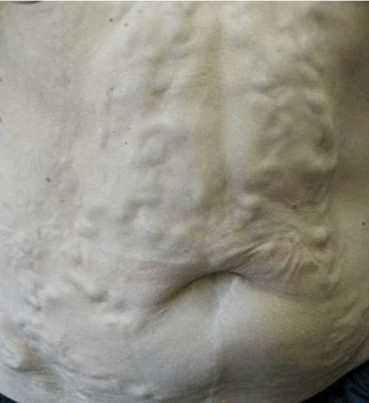
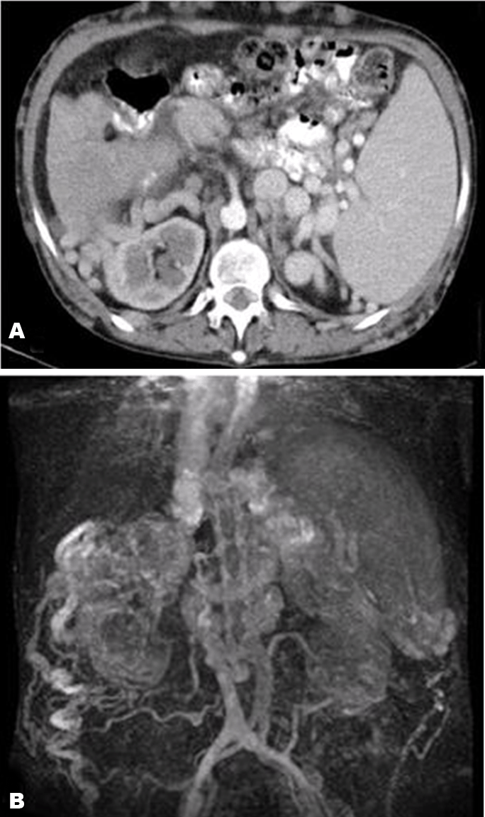
We present a case of 55-year-old female with a history of past alcohol consumption (75 g/day for ten years), with no other relevant medical history, who was referred to our hepatology unit. She presented with leukopenia and thrombocytopenia, normal mean corpuscular volume, mildly elevated total bilirubin (1.8 mg/dL), alkaline phosphatase (192 U/L) and gamma-glutamyl transferase (127 U/L), with normal aminotransferases, albumin and coagulation. Physical examination revealed exuberant collateral circulation of the abdominal wall (Figure 1). The esophagoduodenoscopy revealed grade III esophageal varices, and the abdominal ultrasonography showed hypertrophy of the caudate lobe and heterogeneous echotexture, and splenomegaly with collateral circulation of the splenic hilum. The autoimmune study, hepatitis serologies and iron studies were normal, and the transient elastography was 48 Kpa, consistent with liver cirrhosis. A computed tomography (CT) scan of the abdomen (Figure 2A) revealed exuberant retrocrural collateral circulation, with varices in the azygos and hemiazygos systems, hepatorenal and splenorenal spaces, with lack of permeability in the inferior vena cava, between the renal and the suprahepatic veins. The magnetic resonance angiography (Figure 2B) confirmed inferior vena cava agenesis. Due to beta-blocker intolerance, the esophageal varices were obliterated with endoscopic variceal ligation. She remains in follow-up after three years. | ||||||
| ||||||
|
| ||||||
|
| ||||||
|
Discussion
| ||||||
|
In this case, a history of alcohol consumption had been assumed as cause for liver cirrhosis and evidence of portal hypertension. The exuberance of collateral circulation prompted further image studies, with the diagnosis of inferior vena cava agenesis [1] [2]. Although considered a rare condition, the number of diagnosed cases is increasing, and it is thought that this condition may be underdiagnosed, mainly because most patients are asymptomatic [3]. The best methods for imagiological evaluation are computed tomography scan and magnetic resonance angiography [4]. These individuals generally develop extensive collateral circulation, and it is associated with diverse manifestations, such as deep venous thrombosis [2], lumbosacral radiculopathy and myelopathy due to venous congestion [5], congenital heart disease, splenic abnormalities and liver disease due to inadequate portal flow, with portal hypertension, development of nodal nodular hyperplasia and hepatic tumors. Portal hypertension and hepatic encephalopathy are uncommon before adulthood [4]. The management of inferior vena cava agenesis depends on the anatomical alterations and clinical manifestations of the disease, in a case-by-case basis [4]. High thrombotic risk and presence of liver tumors are important factors to have in mind in patients with this condition. | ||||||
|
Conclusion
| ||||||
|
Inferior vena cava agenesis is a rare condition, but recent evidence shows that it may be underdiagnosed, and can be simply detected with current image methods. Diagnosing inferior vena cava has potential implications on patient management, but it can be challenging due to lack of symptoms. Keywords: Collateral circulation, Inferior vena cava agenesis | ||||||
|
Acknowledgements
| ||||||
|
We are thankful to Sónia Carvalho, MD, for attending of the Hepatology Unit and care provider for the patient | ||||||
|
References
| ||||||
| ||||||

|
[HTML Abstract]
[PDF Full Text]
|
|
Author Contributions
Ana Vaz Cristino – Conception and design, Acquisition of data, Analysis and interpretation of data, Drafting the article, Revising it critically for important intellectual content, Final approval of the version to be published Renata Silva – Acquisition of data, Analysis and interpretation of data, Drafting the article, Revising it critically for important intellectual content, Final approval of the version to be published Carmen Pais – Acquisition of data, Analysis and interpretation of data, Revising it critically for important intellectual content, Final approval of the version to be published José Presa – Acquisition of data, Analysis and interpretation of data, Revising it critically for important intellectual content, Final approval of the version to be published |
|
Guarantor of submission
The corresponding author is the guarantor of submission. |
|
Source of support
None |
|
Conflict of interest
Authors declare no conflict of interest. |
|
Copyright
© 2017 Ana Vaz Cristino et al. This article is distributed under the terms of Creative Commons Attribution License which permits unrestricted use, distribution and reproduction in any medium provided the original author(s) and original publisher are properly credited. Please see the copyright policy on the journal website for more information. |
|
|