
|
 |
|
Case Report
| ||||||
| Obstructed inguinal hernia containing female reproductive organ: A rare presentation | ||||||
| Diyaree N. Ismael1, Zuhair D. Hammood1,2, Fahmi H. Kakamad2,3, Goran A Qadr2,4 | ||||||
|
1Sulaimani Teaching Hospital, Department of Surgery, Sulaimani, Kurdistan
2Kscien Organization for Scientific Research/Hamdi Street, Azadi Building, Sulaimani, Kurdistan 3Faculty of Medical Sciences, School of Medicine, Department Cardiothoracic and Vascular Surgery, University of Sulaimani, Old campus, Sulaimani, Kurdistan 4Faculty of Science & Science Education, School of Science, Biology Department/University of Sulaimani, New campus, Sulaymaniyah, Kurdistan | ||||||
| ||||||
|
[HTML Abstract]
[PDF Full Text]
[Print This Article] [Similar article in Pumed] [Similar article in Google Scholar] 
|


| How to cite this article |
| Ismael DN, Hammood ZD, Kakamad FH, Qadr GA. Obstructed inguinal hernia containing female reproductive organ: A rare presentation. Int J Case Rep Images 2017;8(12):822–825. |
|
ABSTRACT
| ||||||
|
Introduction: Inguinal hernia is a common surgical problem; usually the bowel and omentum herniate through the defect. Herniation of the female reproductive organs is extremely rare. The aim of this study is to report a case of obstructed inguinal hernia with herniation of ovary and uterine tube. Keywords: Hernia, Ovary, Uterine tube | ||||||
|
INTRODUCTION
| ||||||
|
The inguinal hernia is a relatively common surgical problem in kids with an accounted occurrence of 0.8–4.4% [1]. About 13.7–23% of inguinal hernias of indirect type happen in young females [2]. The proportion of male to young ladies is 6:1.1. Nearly, 68.1% occur on the right side, 23.4% on the left side and 8.5% are bilateral. The accounted frequency is about 71% for youngsters under five years and 30% for youths or ladies in reproductive ages [3]. The most common organs involved in the hernia pathology are omentum and small bowel. Other intra-abdominal organs are infrequently encountered in the inguinal canal such as the vermiform appendix, urinary bladder and fibroid. Very rarely, ovaries and fallopian tubes have been reported as content of inguinal hernia sac which presented challenge to the physician and surgeon [4]. The inguinal hernia containing a reproductive organ in young woman usually happens due to a partial closure of the peritoneal processus vaginalis. Unfortunately, the repair of hernia in female child is usually underwent with much less care than in male because of absence of spermatic cord in female, this led to different type of injuries to both ovary and uterine tube [4][5]. The aim of this report is to discuss an uncommon case of left obstructed sliding inguinal hernia with left ovary containing a large ovarian cyst along with it is uterine tube. | ||||||
|
CASE REPORT
| ||||||
|
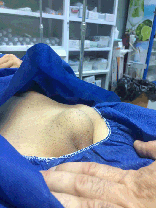
A 14-year-old girl a student of secondary school and known case of neglected, reducible left inguinal hernia since two years ago, was admitted to the emergency room with a two-day history of tender irreducible swelling of left groin associated with nausea but no vomiting. On examination; There was 5x6 cm, irreducible, tender left inguinal swelling (Figure 1), the abdomen was soft and the bowel sound was positive. Vital signs were stable apart from tachycardia (114 beats per minute). Urgent ultrasound examination was done which proved the diagnosis as irreducible left inguinal hernia containing left ovary with complex cystic content. After informed consent, she was taken to the emergency operating room. Under general anesthesia with endotracheal tube and single prophylactic antibiotic, exploration of the left inguinal canal was done. The hernia sac and its contents were isolated which were irreducible below the superficial inguinal ring. The hernia was of indirect type and the contents were ovary containing large cyst and the fallopian tube. The sac was opened; both ovary and fallopian tube were viable while the fimbrias were gangrenous (Figure 2). The ovarian cyst was excised and the small gangrenous part of the fimbrias was left, the sac closed then reduced to the abdominal cavity. Round ligament was preserved and the posterior wall was reinforced by Darning procedure (Figure 3). The wound was closed in layers and the patient was kept in hospital for two days. Postoperative period was uneventful. The skin stitches were removed one week later. | ||||||
| ||||||
| ||||||
| ||||||
|
DISCUSSION
| ||||||
|
In female, the inguinal canal normally forms a passage for the round ligament of the uterus, processus vaginalis and labial arteries [5]. The round ligament formed by the distal part of the gubernaculums, while proximal part forms the suspensory ligament of the ovary [1]. The reproductive systems of both females and males share the same steps in the early uterine development. Understanding this fact may help understanding the mechanism of the ovarian herniation through the inguinal canal [6]. The pathophysiology of the development of sliding inguinal hernias of ovary and fallopian tube in female is thought to be a homologous to the normal physiology of the testis descent in male [1]. Increased pressure over the hernia may compromise blood supply of its contents, especially venous one, and may lead to venous congestion and subsequently ischemia and infarction. Another risk of vascular compromise is torsion of the ovary as it has a long pedicle [2]. In this case, both the ovary and the tube were viable. Differential diagnoses of the inguinal region swelling in the female are varied, which may include direct inguinal hernia, indirect inguinal hernia, soft tissue tumors (sarcoma, leiomyoma or lipoma), cystic lesions, abscess collection, enlarged lymph nodes, or hydrocele [1]. The diagnosis can be easily confirmed by ultrasonography scan in most cases. However, for better anatomical illustration and details of the hernias and its boundaries as well as contents magnetic resonance imaging (MRI) and computed tomography (CT) scan can be done [7]. The current case was successfully diagnosed preoperatively by ultrasound. When the inguinal hernia sac contents formed by ovary and uterine tube, they are usually associated with developmental anomalies of the genital tract like bicornuate uterus, vaginal atresia and renal anomalies. The current case was free from other anomalies [8]. Inguinal hernia is treated with reduction of its content provided that there is no abnormality of the ovary or the fallopian tube, no impaired blood supply and there are no features of salpingitis. Reduction of the contents should be followed with ligation at high level of the sac then closure of deep inguinal ring and finally re-supporting of the posterior wall of the inguinal canal by a non-absorbable mesh in females aged more than 20 years, the procedure which is performed in this case [8]. Associated injury to the hernia content is a common problem especially in females with sliding inguinal herniation of vital reproductive organs like ovaries, fallopian tubes and uterus. Therefore, ligation of the hernia sac without opening should be avoided. However, in all cases with inguinal hernia, the sac should be opened during repair to exclude the presence of sliding ovaries or fallopian tubes, and if present, should be dissected and reduced carefully to abdominal cavity before high ligation of the sac to avoid their associated injuries [1]. In current case, the herniated ovary and uterine tube was safely reduced. | ||||||
|
CONCLUSION
| ||||||
|
Inguinal hernia with ovarian and uterine tube herniation is a very rare disease, when it present, it can be diagnosed by ultrasonography, and management is by surgery with careful reduction of the content. | ||||||
|
REFERENCES
| ||||||
| ||||||


|
[HTML Abstract]
[PDF Full Text]
|
|
Author Contributions
Diyaree N. Ismael – Substantial contributions to conception and design, Acquisition of data, Analysis and interpretation of data, Revising it critically for important intellectual content, Final approval of the version to be published Zuhair D. Hammood – Substantial contributions to conception and design, Drafting the article, Revising it critically for important intellectual content, Final approval of the version to be published Fahmi H. Kakamad – Substantial contributions to conception and design, Drafting the article, Revising it critically for important intellectual content, Final approval of the version to be published Goran A. Qadr – Substantial contributions to conception and design, Drafting the article, Final approval of the version to be published |
|
Guarantor of Submission
The corresponding author is the guarantor of submission. |
|
Source of Support
None |
|
Conflict of Interest
Authors declare no conflict of interest. |
|
Copyright
© 2017 Diyaree N. Ismael et al. This article is distributed under the terms of Creative Commons Attribution License which permits unrestricted use, distribution and reproduction in any medium provided the original author(s) and original publisher are properly credited. Please see the copyright policy on the journal website for more information. |
|
|