|
|
|
|
Case Report
| ||||||
| A case of tuberculosis in the appearance of a metastatic tumor | ||||||
| Yilmaz Baş1, Ali Bulgan2, Ikram Abdikarim Ibrahim3 | ||||||
|
1Department of Pathology, Hitit University Training and Research Hospital, Çorum, Turkey
2Department of Radiology, Iskenderun State Hospital, Hatay, Turkey 3General Surgery, Somali Mogadishu-Turkey Training and Research Hospital, Mogadishu, Somali | ||||||
| ||||||
|
[HTML Abstract]
[PDF Full Text]
[Print This Article] [Similar article in Pumed] [Similar article in Google Scholar] 
|
| How to cite this article |
| Baş Y, Bulgan A, Ibrahim IA. A case of tuberculosis in the appearance of a metastatic tumor. Int J Case Rep Images 2017;8(11):698–702. |
|
ABSTRACT
| ||||||
|
Introduction: We report a case of chronic osteolysis of the ribs, iliac bones and vertebrae, Pott’s abscess due to tuberculosis that was previously misdiagnosed as cancer metastasis on computed tomography imaging. Keywords: Bone and joint infections, Pathology, Respiratory infections, Tuberculosis | ||||||
|
INTRODUCTION
| ||||||
|
Tuberculosis remains a world-wide problem for more than a century ago, and it affects all age groups [1]. In Somali Regional State, Eastern Ethiopia, a total of 31, 198 all types of new tuberculosis cases were registered and reported from 2003–2012 had a total population of 4, 445, 219 [2], and the estimated incidence rate in Sub-Saharan Africa is also nearly 700 cases per 100,000 populations [3]. In 2014, the World Health Organization (WHO) identified nine million new cases of the disease with 1.5 million deaths [1]. Two to five percent of all tuberculosis in the world and 11–15% of extrapulmonary tuberculosis are osteoarticular tuberculosis [1]. Extrapulmonary tuberculosis rates reported respectively lymphatic (42.0%), pleural (18.0%), bone and/or joint (11.1%), meningeal (6.3%), peritoneal (5.5%), genitourinary (5.2%), other (11.9%) in the United States, 2005 [4]. Vertebra-localized tuberculosis is known as Pott’s disease and psoas abscesses are suppurative collections within the fascia surrounding the psoas and iliacus muscles [3][5]. Clinically and radiologically, it can be confused with cancer metastatic lesions as a result of multifocal bone involvement [6]. The diagnosis of such cases is made by evaluating the clinical, microbiological, radiological and histopathological findings collectively [5][6]. Histopathological findings are very important in diagnosis [5]. A case of multiple bone lesions and with psoas abscess, presented in the light of literature because of its clinico-radiological presentation as a cancer metastasis. | ||||||
|
CASE REPORT
| ||||||
|
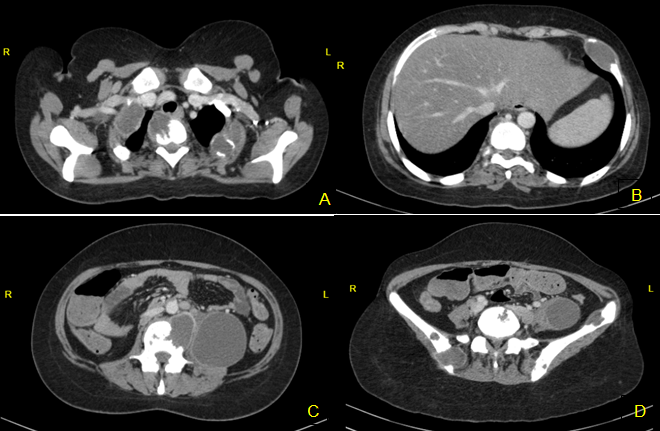
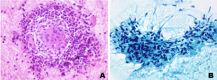
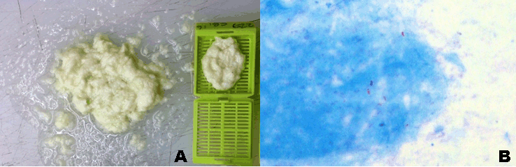
A 28-year-old Somali female was admitted to the Somali Mogadishu-Turkey Training and Research Hospital general surgery service for abdominal pain, subcutaneous left anterior chest wall swelling and sensitivity, lumbovertebral sensitivity, backache, severely pale, sweating, fever, and weight loss. Historically, she had a negative history of contact with active tuberculous and not received the Bacillus Calmette-Guérin (BCG) vaccine. Patient’s family had never been diagnosed with tuberculosis. She was not diabetic or immunosuppressed and she denied any trauma to the site. Physical examination revealed swelling of the tissue overlying the ribs. The vertebral-spines, ribs area and left thigh were firm and painful to touch. And she had moderate kyphosis. There was no BCG scar. Computed tomography (CT) scan of the chest, abdomen and iliac bone confirmed multiple expansive, destructive hypodense mass or abscess lesion on the right 1st, 2nd, 4th ribs, the left 1st, 2nd, 6th ribs and 1st, 2nd, 6th, 10th, 12th thoracic vertebrae, 1st, 2nd, 3th, 4th lumbar vertebrae, the left and the right iliac bones (Figure 1A–D). Additionally, there was a 8x7 cm hypodense area compatible with abscess or hematoma on the left psoas muscle and along the left iliopsoas muscle. Laboratory tests of the patient showed leukocytosis at 15,000/mL, erythrocyte sedimentation rate (ESR) 65 mm, CRP 38 mg/l and CEA 1.5 ng/ml. Other laboratory tests were within normal limits. No tuberculin skin test was performed. HIV and Brucella serology was negative. A fine needle aspiration biopsy (FNAB) of the 6th rib lesion was performed. Cytopathological examination revealed an epithelioid and giant cell granulomatous inflammatory process consistent with tuberculosis (Figure 2). An ultrasonography (US) guided percutaneous drainage of the abscess was performed. Purulent cream colored liquid was drained (Figure 3A). Slides of cell block were prepared from the drainage material. The Ziehl–Neelsen staining of aspiration was positive (Figure 3B). | ||||||
|
| ||||||
|
| ||||||
| ||||||
|
DISCUSSION
| ||||||
|
Tuberculosis is a disease that remains endemic in many parts of the world particularly in developing countries [1]. Immune deficiency, low socio-economic level, dialysis, transplantation, malignancy pathology, infants, and malnutrition are essential risk factors [1][5]. In addition, there are populations who do not have access to BCG vaccine in regions such as Somalia. In our case, low socio-economic status factor was present and also did not have BCG scar. In countries such as Somali, where tuberculosis is endemic, and the diagnosis is traditionally made on clinical and radiological grounds. Histopathological confirmation is not usually undertaken, because it is expensive and the cytopathology department has not yet settled in many centers in spite of there are the large case load. Therefore, we performed FNAB at the lesion in the rib because of our pathology department in Somali Magodishu-Türk Hospital has enough facility. In the study of S Bhojraj et al. [7] biopsy for pathologic examination was not attempted in patients with osteitis. Most of their patients (93%) were diagnosed and treated satisfactorily on the basis of clinical and radiological evidence, and without histopathological diagnosis. According to them study, a therapeutic trial of antituberculous treatment is a practical alternative to taking a biopsy [7]. But as noted in Urbanczik’s study [5] in bone samples from areas other than the spine, histopathology remains the gold standard technique disclosing the classical caseating tubercle granulomas. Our case was diagnosed according to radiological and cytomorphological findings. Total 14,097 tuberculosis cases reported to Centers for Disease Control (CDC) in 2005 [4]. There were 329 (11.1%) patients with bone and/or joint involvement, and ranks third after the pleural and lymphatic tuberculosis [4]. In study of Lemnouer et. al. [1], osteoarticular tuberculosis has found the fourth most prevalent tuberculosis localization after pulmonary, urogenital and lymphatic. In our case, the patient had multiple expansive, destructive lytic, mass or abscess lesion vertebrae, ribs, iliac bones with psoas abscess. The lung and other organ involvement were not observed. Tuberculosis is second only to metastatic neoplasms as a cause of destructive multifocal bone lesions such as ribs lesions [6]. Typhoid fever or paratyphoid fever, actinomycosis, syphilis, infections due to streptococci or staphylococci, coccidioidomycosis, blastomycosis, and brucellosis are other infectious etiologies [6]. Malignancies include metastases from primary carcinoma of the liver, breast, thyroid, and kidney; Ewing’s sarcoma; fibrosarcoma; multiple myeloma; or histiocytosis [6][8]. Cytomorphological examination revealed no malignant cells in our case, and Brucella serology was negative. Vertebral tuberculosis, which has two different patterns, is a chronic, slow-progressive disease [8]. The first is spondylitis without disc involvement, which is exceedingly more common, and multilevel vertebral body involvement could observe in this form as skip lesions [8]. The second pattern is destruction of two or more contiguous vertebrae associated with late-onset disc infection, which results in intervertebral space narrowing due to disc herniation into the collapsed vertebral body [8]. Spinal spondylitis is the most common manifestation of osteoarticular tuberculosis, and 1–3% of patients with tuberculosis have skeletal involvement [9]. The lumbar spine is the most common site of the disease followed by the thoracic region [3]. In metastatic disease, thoracic region is most commonly involved, while posterior wall of the vertebral body (60%), pedicles and lamina (50%) are involved. However, intervertebral disc heights are preserved [3]. In multiple vertebral body involvement, the characteristic gibbous deformity and paraspinal mass or collection are common. Posterior element involvement is a characteristic of vertebral tuberculosis [8]. In this case, the patient has lesions at T2nd, T10th, T12th, L1st, L2nd, L3th corpuses, T5th right pedincule, L1st left pedincule and spinous process, L4th left pedincule expansive, destructive lytic, mass or abscess lesion. When severe, distinct loss of vertebral height can lead to moderate kyphosis with facet joint subluxation or dislocation. Preservation of disc space as well as posterior element abnormalities and multiplicity of vertebral body involvement in tuberculosis may lead to difficulty in distinguishing tuberclusis from tumoral processes. Early symptoms of vertebral tuberculosis are relatively nonspecific, and include backache, vertebral sensitivity, fever, and weight loss [8]. In the patient, there was backache, sweating, weight loss, tenderness in ribs and vertebrae. She had moderate kyphosis. Tuberculosis of the ilium constitutes less than 1% of all skeletal tuberculosis [8]. A typical radiological feature of tuberculosis in cancellous bones is feathery sequestra or lytic lesions with irregular margins associated with mild surrounding sclerosis [8]. In our patient has multiple expansive, destructive lesions on the left and the right iliac bones. Tuberculosis of the rib occurs in males about twice than females [6]. This infection occurs most often in patients between 15 and 30 years of age which occurs most often in children between the ages of 2 and 10 years [6]. Our patient was a 28-year-old female. Rib involvement within skeleton tuberculosis is at 1.7%, however, it is the second widespread disease next to metastatic lesions among the destructive lesions of the ribs [10]. Hence, physical and radiological examination findings can often be confused with primary and metastatic chest wall tumors [6]. Rib tuberculosis frequently emerges until 18 months after infection, and <50% of patients have active pulmonary disease [6][10]. Therapy is based on 9–12 months of antituberculous chemotherapy [1]. The disease may be drug-resistant, however, and therefore a poor response to standard hemotherapy at the end of 6–12 weeks does not imply that surgery is indicated [7]. In our case, the patient has multiple expansive, destructive mass or abscess lesion on the right 1st, 2nd, 4th ribs and on the left 1st, 2nd, 6th ribs. Likewise, tuberculosis cases with rib involvement with Pott’s disease are rare too [7]. Iliopsoas abscess may be classified as primary or secondary, and primary iliopsoas abscess occurs probably as a result of hematogenous spread of an infectious process from an occult source in the body [11]. Crohn’s disease, urinary tract infections, vertebral osteomyelitis, infected aortic aneurysm, endocarditis, intrauterine contraceptive device, suppurative lymphadenitis etc. are also associated with secondary to iliopsoas abscess [11]. Treatment involves the use of appropriate antibiotics along with drainage of the abscess [11]. Drainage of an abscess in iliopsoas compartments may be indicated if it causes symptoms which are simply due to its size and location, such as a flexion deformity of the hip [7]. A neurological deficit is not an absolute indication for surgery, and responds well to conservative treatment [7]. However, the mortality rate increases with delayed and incorrect diagnoses and reaches 100% in cases without drainage [12]. Our patient denied any trauma and had an abscess formation, starting from the T12 vertebra and extending up to thigh. The case in light of these findings has been investigated as a secondary Pott’s disease. Abscess drainage was performed and tuberculosis treatment was started. There was no neurological deficit on physical examination. But, as noted in the study of Woldeyohannes et al. [2] treatment completion rate in Somalia was relatively lower than the value in the world, Europe, Turkey and report from Arsi Zone, Central Ethiopia. | ||||||
|
CONCLUSION
| ||||||
|
To conclude, there are population that do not have access to BCG vaccine in these regions such as Somali. We have shown the clinico-radiological findings of a case with multifocal osseous, initially misdiagnosed as cancer metastases. Familiarity with the imaging features of musculoskeletal tuberculosis and a high index of clinical suspicion are necessary for the correct diagnosis and proper treatment. Histopathological findings are also very important in diagnosis. | ||||||
|
REFERENCES
| ||||||
| ||||||
|
[HTML Abstract]
[PDF Full Text]
|
|
Author Contributions
Yilmaz Baş – Substantial contributions to conception and design, Acquisition of data, Analysis and interpretation of data, Drafting the article, Revising it critically for important intellectual content, Final approval of the version to be published Ali Bulgan – Analysis and interpretation of data, Revising it critically for important intellectual content, Final approval of the version to be published Ikram Abdikarim Ibrahim – Analysis and interpretation of data, Revising it critically for important intellectual content, Final approval of the version to be published |
|
Guarantor
The corresponding author is the guarantor of submission. |
|
Source of support
None |
|
Conflict of interest
Authors declare no conflict of interest. |
|
Copyright
© 2017 Yilmaz Baş et al. This article is distributed under the terms of Creative Commons Attribution License which permits unrestricted use, distribution and reproduction in any medium provided the original author(s) and original publisher are properly credited. Please see the copyright policy on the journal website for more information. |
|
|