|
|
|
|
Clinical Image
| ||||||
| Complete spontaneous resolution of vanishing lung syndrome: A rare case | ||||||
| Roy Cho1, Felix D. Zamora1, Heidi H. Gibson2, Erhan Dincer3 | ||||||
|
1MD, Assistant Professor, University of Minnesota, Department of Pulmonary, Allergy, Critical Care and Sleep Medicine 2RRT, Lead-interventional Respiratory Therapist, Cardiopulmonary Services, University of Minnesota 3MD, Associate Professor, University of Minnesota, Department of Pulmonary, Allergy, Critical Care and Sleep Medicine | ||||||
| ||||||
|
[HTML Abstract]
[PDF Full Text]
[Print This Article]
[Similar article in Pumed] [Similar article in Google Scholar] 
|
| How to cite this article |
| Cho R, Zamora FD, Gibson HH, Dincer E. Complete spontaneous resolution of vanishing lung syndrome: A rare case. Int J Case Rep Images 2017;8():756–757. |
|
CASE REPORT
|
|
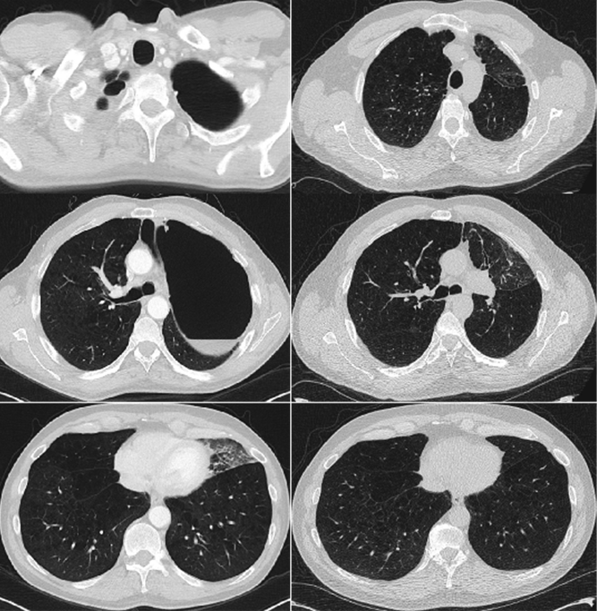
A 59-year-old male with chronic obstructive pulmonary disease (COPD) returned to the pulmonary clinic three-years after initial evaluation for bronchoscopic lung volume reduction for a giant left upper lobe bullae. He had moderately severe COPD (FEV1 1.6L, 37%) controlled on inhaler therapy and a greater than 35-pack/year smoking history. Previously, he was deemed a candidate for lung volume reduction. However, declined and was lost to follow-up (Figure 1). On presentation, he denied any decline in his physical capacity for the past three-years. His repeat chest CT scan demonstrated complete resolution of the large left upper lobe bullae. As a result of these findings, we did not recommend any further intervention with regard to the previous bullae. However, recommended annual lung cancer surveillance with low-dose chest CT scan. |
|
|
|
DISCUSSION
|
|
Giant pulmonary bullae or vanishing lung syndrome (VLS) is defined by radiographic criteria including presence of giant bullae in one or both upper lobes, occupying at least one-third of the hemithorax and compressing surrounding normal lung parenchyma [1]. The natural history of VLS is unpredictable and is based on case reports and experience forming expert opinion. The leading theory for bullae expansion is air trapping that impedes expiratory air-flow leading to tension and gradual enlargement of the air spaces [2]. Although most bullae enlarge, only three-case reports since 1990 have shown regression of the disease [3][4][5]. These cases report partial regression following instances of airway obstruction (infection, inflammation, etc.); in which, the leading hypothesis is complete isolation of the space leading to shrinkage via air and fluid resorption over time. Notably, this is the first case to demonstrate complete resolution of VLS radiographically. |
|
CONCLUSION
|
|
Usual course for vanishing lung syndrome (VLS) is progression of disease and worsening pulmonary function as measured by FEV1. However, we report a very rare case of VLS that has completely resolved without any intervention. Keywords: Emphysematous bullae, Interventional pulmonary, Vanishing lung syndrome |
|
REFERENCES
|
|
|
[HTML Abstract]
[PDF Full Text]
|
|
Author Contributions
Roy Cho – Substantial contributions to conception and design, Drafting the article, Revising it critically for important intellectual content, Final approval of the version to be published Felix D. Zamora – Substantial contributions to conception and design, Drafting the article, Final approval of the version to be published Heidi H. Gibson – Substantial contributions to conception and design, Drafting the article, Final approval of the version to be published Erhan Dincer – Substantial contributions to conception and design, Drafting the article, Final approval of the version to be published |
|
Guarantor
The corresponding author is the guarantor of submission. |
|
Source of support
None |
|
Conflict of interest
Authors declare no conflict of interest. |
|
Copyright
© 2017 Roy Cho et al. This article is distributed under the terms of Creative Commons Attribution License which permits unrestricted use, distribution and reproduction in any medium provided the original author(s) and original publisher are properly credited. Please see the copyright policy on the journal website for more information. |
|
|