|
 |
|
Case Report
| ||||||
| Percutaneous drainage of delayed traumatic subcapsular hematoma of the spleen following splenic salvage: A case report | ||||||
| Alaa Sedik1, Mahmood Makhdoomi2, Abrar Hussein3, Salwa Elhoushy4, Ahmed Morsy5 | ||||||
|
1MBChB, MS, FRCS (Glasg), DMAS (Delhi), Consultant General Surgeon, King Khalid Hospital, Hail, Saudi Arabia 2Specialist General Surgeon, King Khalid Hospital, Hail, Surgery Department, Saudi Arabia 3General Surgery Resident, King Khalid Hospital, Hail, Saudi Arabia 4MBChB, MRCP (UK), Medical Specialist, King Khalid Hospital, Hail, Medical Department, Saudi Arabia 5Radiological Specialist, King Khalid Hospital, Hail, Saudi Arabiac | ||||||
| ||||||
|
[HTML Abstract]
[PDF Full Text]
[Print This Article] [Similar article in Pumed] [Similar article in Google Scholar] 
|


| How to cite this article |
| Sedik A, Makhdoomi M, Hussein A, Elhoushy S, Morsy A. Percutaneous drainage of delayed traumatic subcapsular hematoma of the spleen following splenic salvage: A case report. Int J Case Rep Images 2017;8(10):639–642. |
|
ABSTRACT
|
|
Introduction: Percutaneous image-guided splenic procedures are seldom performed due to fear of complications, mainly hemorrhage. The reported cases in literature were due to atraumatic causes. These procedures obviate the need for splenectomy. Thus, preserving the spleen and decrease the pressure to avoid possible rupture. Keywords: Hematoma, Percutaneous drainage, Spleen |
|
INTRODUCTION
|
|
Splenic salvage is less commonly practiced nowadays. Delayed splenic rupture is mostly due to ruptured hematoma and carries higher mortality (5–15%) than acute (1%) [1][2][3] . The management of non-traumatic subcapsular splenic hematoma remains controversial. Splenectomy or percutaneous drainage under ultrasound guidance may be done. No reports are available in the literature on percutaneous drainage of traumatic hematoma. We herein reported a 30-year-old male, a victim of road traffic accident (RTA), with large traumatic subcapsular splenic hematoma after splenic salvage that was managed successfully by percutaneous ultrasound-guided drainage. |
|
CASE REPORT
|
|
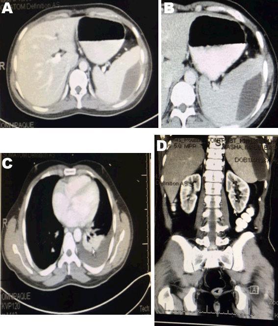
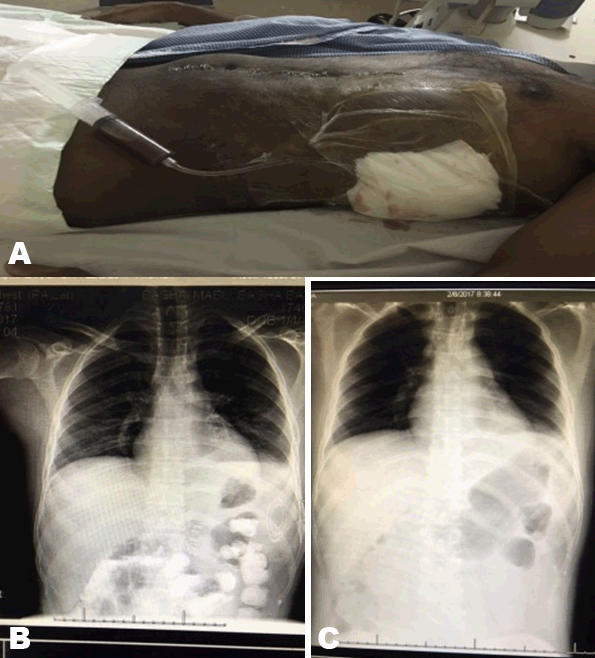
A 30-year-old Indian male presented to our emergency room, a RTA victim, with lower abdominal pain. Resuscitation started according to ATLS protocol and examination was unremarkable except for suprapubic tenderness, tear of glans penis and right side of scrotum. Laboratory workup showed raised renal functions. Focused assessment with sonography for trauma (FAST) showed mild free intraperitoneal fluid. X-rays showed bilateral lung contusions and pelvic fracture. Computed tomography (CT) scan without contrast was not done due to raised serum creatinine of 145 µmol/L and urea of 11 mmol urea per liter. Scrotal ultrasonography and Doppler were normal. Repeated hemoglobin was 8 g/dl, then ultrasound showed increased intra-abdominal fluid. He was prepared for laparotomy. 1.2 liters of free blood, normal solid organs except for 1 cm lateral capsular splenic tear with minimal bleeding were found. Splenic salvage was done with cautery and 2 pieces of pieces of local hemostats (Surgicel™). Resection anastomosis of 30 cm of ilium was done for transverse large mesenteric tear of the distal ileum. Wash with saline then, the abdomen was closed over drains. On the next day, he developed hematuria. Computed tomography (CT) scan with contrast showed normal solid organs. On day-10, blood workup and ultrasound were unremarkable. Drains removed and antibiotics were discontinued. He remained for wound infection care. On day-18, he had fever and subcostal pain. Clinically, he had mild left subcostal tenderness with no masses. Ultrasonography and abdominal contrast enhanced CT scan showed a subcapsular 12x6.5x3.6 cm splenic hematoma with no free intraperitoneal fluid, left pleural effusion and basal lung consolidation (Figure 1). Conservative treatment continued for 10 days and till complaining of pain. Follow-up CT scan showed no improvement. He was offered ultrasound-guided percutaneous drainage or splenectomy as alternative in case of failure of drain. 40 ml of turbid blood was aspirated and sent for culture. A 16 F pigtail catheter was left inside the cavity. Post procedure chest X-ray showed a left hydropneumothorax that improved later (Figure 2). The culture of the aspirate was negative. He did well. Regular catheter care with flushing with 5 ml saline. On day-9, the drainage was reduced to 5 ml and serous. The catheter drainage stopped after three days. Ultrasonography showed obliteration of the cavity. The catheter was removed. He was discharged in a good condition and came for outpatient department follow-up three weeks later and was free of complaints. He did not agree to do follow-up CT scan as he had it three times during the course of treatment. CBC and ultrasonography were normal. He has regular follow-up. |
|
|
|
|
|
DISCUSSION
|
|
The spleen is one of the most commonly injured intra-abdominal organs mostly due to blunt trauma [1]. The preservation of functional splenic tissue is secondary and in selected patients may be accomplished using non-operative management with or without splenic angioembolization or operative salvage techniques. Splenectomy remains a life-saving measure for many patients and may be a part of damage control surgery [2]. The American Association for the Surgery of Trauma classified splenic injuries using CT scan [3][4]. The decision to perform splenectomy versus splenic salvage (i.e., splenorrhaphy, partial splenectomy, electrocautery or topical hemostats or argon beam) is based upon the grade of injury, presence of associated injuries, patient’s overall condition, and experience of the surgeon. Our patient was stable intraoperatively and had a low grade(I), then a salvage with cautery of the bleeding point and topical hemostat done. Splenic salvage was extensively practiced before. Stable patients with lower grades are managed non-operatively. Failure will require splenectomy. All these factors has made salvage a rare procedure and the surgeon experience has declined [5][6]. Percutaneous drainage of splenic subcapsular collections may be a feasible and successful treatment, but there are only few reports available in literature. They discussed the atraumatic and spontaneous hematomas complicating mostly acute or chronic pancreatitis [7][8][9]. To the best of our knowledge, this is the first case reported in the literature with a traumatic splenic subcapsular hematoma treated successfully with this modality that needs further investigation and work in future. |
|
CONCLUSION
|
|
The definitive management of subcapsular splenic hematoma complicating trauma is not yet established. Surgery is the treatment of choice for hemodynamically unstable patients. Follow-up of patients with splenic injury treated non-operatively or after savage; after discharge may detect a serious complication and may avoid delayed rupture. Image-guided percutaneous drainage appears to be another feasible option for large subcapsular traumatic splenic hematomas to prevent splenic rupture and obviate the need for splenectomy. |
|
REFERENCES
|
|

|
[HTML Abstract]
[PDF Full Text]
|
|
Author Contributions
Alaa Sedik – Substantial contributions to conception and design, Acquisition of data, Analysis and interpretation of data, Drafting the article, Revising it critically for important intellectual content, Final approval of the version to be published Mahmood Makhdoomi – Acquisition of data, Analysis and interpretation of data, Revising it critically for important intellectual content, Final approval of the version to be published Abrar Hussein – Acquisition of data, Analysis and interpretation of data, Drafting the article, Final approval of the version to be published Salwa Elhoushy – Analysis and interpretation of data, Drafting the article, Revising it critically for important intellectual content, Final approval of the version to be published Ahmed Morsy – Acquisition of data, Analysis and interpretation of data, Revising it critically for important intellectual content, Final approval of the version to be published |
|
Guarantor
The corresponding author is the guarantor of submission. |
|
Source of support
None |
|
Conflict of interest
Authors declare no conflict of interest. |
|
Copyright
© 2017 Alaa Sedik et al. This article is distributed under the terms of Creative Commons Attribution License which permits unrestricted use, distribution and reproduction in any medium provided the original author(s) and original publisher are properly credited. Please see the copyright policy on the journal website for more information. |
|
ABOUT THE AUTHORS
| |||||||||||||||
| |||||||||||||||
|
|