|
 |
|
Letter to the Editor
| ||||||
| Common peroneal nerve palsy caused by an initially misdiagnosed extraneural and intraneural benign ganglion cyst of the peroneal nerve in a 11-year-old child: A rare but severe condition | ||||||
| Ingo Schmidt | ||||||
|
SRH Poliklinik Gera GmbH, Straße des Friedens 122, 07548 Gera (Germany)
| ||||||
| ||||||
|
[HTML Abstract]
[PDF Full Text]
[Print This Article] [Similar article in Pumed] [Similar article in Google Scholar] 
|


| How to cite this article |
| Schmidt I. Common peroneal nerve palsy caused by an initially misdiagnosed extraneural and intraneural benign ganglion cyst of the peroneal nerve in a 11-year-old child: A rare but severe condition. Int J Case Rep Images 2017;8(9):623–626. |
| To the Editor, |
|
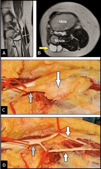
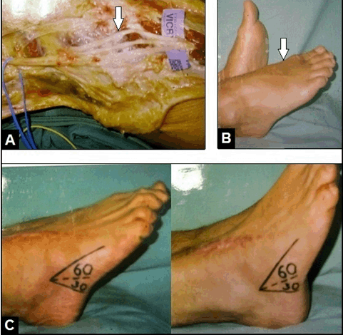
An 11-year-old obese boy presented with a right common peroneal nerve (CPN) palsy. At first presentation in our hospital, the patient reported progressive palsy starting only with painless weakness for dorsiflexion of his right foot two years ago. There was no history of any trauma. Within these two years, the patient was explored by his family doctor, a pediatrics, and a neurologist. The diagnostic management included magnetic resonance imaging (MRI) of the cerebrum and overall spine, electromyography, electroencephalography, analyses of blood and cerebrospinal fluid samples. No causes could be found by the treating physicians that declared his CPN palsy. So the diagnosis of a peroneal nerve mononeuropathy with unclear genesis was made, and the patient was treated by oral medication of glucocorticoids and vitamin B12. On first clinical examination in our hospital, a painless foot drop was present, and the sensibility at the peripheral peroneal nerve area was completely lost. The strength of dorsiflexion of foot was completely lost according grade 0 in Medical Research Council scale (0–5), and electromyography revealed a severe axonal lesion in the absence of motor activities. Magnetic resonance imaging (MRI) scans of his right knee revealed a subcutaneous, well demarcated and lobulated cyst adjacent to the proximal tibiofibular joint which was clinically not palpable through the thick layer of subcutaneous fat of the obese patient (Figure 1A–B). According to these findings, the diagnosis of a proximal tibiofibular joint cyst was made by the radiologist. Based on the presented peripheral neurological symptoms, surgical revision was detected by us. The lesion was surgically exposed through a large longitudinal incision starting distally over the lateral aspect of fibula, passing the popliteal fossa with a vertical incision, and extending up longitudinally on the dorsal aspect of the thigh. Intraoperatively, a cyst originating from the proximal tibiofibular joint could not be found, but there was an extraneural and intraneural ganglion cyst with size of 7x2.5 cm involving the CPN, and both its superficial sensory nerve (SSN) and deep motor nerve (DMN) distally (Figure 1C). After careful dissection and removal of the tumor in a monobloc manner, a division of the CPN distally was seen, but macroscopically there were no structural damages (i.e., neurotmesis) both of the CPN and its DMN/SSN (Figure 1D). Histological examination confirmed the diagnosis of a benign ganglion cyst. The wound healing was uneventful. One year after surgery, a functional recovery could not be observed, so a tendon transfer procedure to restore dorsiflexion of his foot was recommended by us, but declined by the parents of the patient at this time. The CPN originates from the sciatic nerve, passes posteriorly and laterally to the biceps femoris muscle in the popliteal fossa, crosses the head of the fibula, and descends to lower leg after dividing in its SSN and DMN. Most often, CPN palsy occurs at the fibular neck, where the nerve is superficial and vulnerable to direct injury, overstretching, and entrapment. The entrapment of CPN potentially leading to palsy (i.e., foot drop) caused by extraneural tumors around the proximal tibiofibular joint is known from literature [1]. Nerve entrapment caused by ganglion cysts is still widely known from the upper extremity. Ganglion cysts around the proximal tibiofibular joint leading to CPN palsy, first described in 1921 by Sultan [2], is much less common, it has a peak incidence in the fourth decade of life, and it may occur both extraneural and intraneural [3][4][5]. Our patient’s case suggested that ganglion cysts of the peroneal nerve in MRI can mimicking proximal tibiofibular joint cysts. Its prevalence with 0.76% is also very rare that was found in a cross-sectional study including 654 knee MRI scans in patients with the most common clinical diagnosis of meniscal tears (42.8%), the mean age of these patients was 43.4 years (range 42–54 years), and the cysts ranged in size from 1.0–2.8 cm [6]. In literature, some case reports have been described CPN palsy caused by a ganglion cysts of the peroneal nerve or a cyst originating from the proximal tibiofibular joint in children [7][8][9][10][11][12]. With our patient, it must be noted critically that the ganglion cyst was initially misdiagnosed over a period of two years, and so a functional recovery could not be achieved, despite surgical removal of the cyst and neurolysis of the CPN. It must be suggested that the prolonged time in diagnostic management (initially demyelinating entrapment with weakness only) led to a severe and irreparable axonotmesis of the peroneal nerve. When a CPN palsy is present, surgical nerve decompression with neurolysis should be done as early as possible. The prognosis of functional recovery in patients with CPN palsy depends on the severity of neurological deficit and time of surgical neurolysis. In general, the prognosis for a demyelinating lesion is much more favorable than for an axonal loss lesion [13]. The outcome is usually favorable if surgery is done within four months after first presentation of neurological deficits, and less favorable in patients who have neurological symptoms for longer than one year [1][14]. For irreparable CPN palsy, a tendon transfer procedure utilizing the tibialis posterior, first described in 1933 by Ober [15], with or without a combined transfer of the flexor digitorum longus is the method of choice for functional recovery of patients (Figure 2A–C) [16][17]. |
|
|
|
|
|
Keywords:Ganglion cyst peroneal nerve, Common peroneal nerve palsy |
|
References
|
|

|
[HTML Abstract]
[PDF Full Text]
|
|
Author Contributions
Ingo Schmidt – Substantial contributions to conception and design, Acquisition of data, Analysis and interpretation of data, Drafting the article, Revising it critically for important intellectual content, Final approval of the version to be published |
|
Guarantor of submission
The corresponding author is the guarantor of submission. |
|
Source of support
None |
|
Conflict of interest
Authors declare no conflict of interest. |
|
Copyright
© 2017 Ingo Schmidt. This article is distributed under the terms of Creative Commons Attribution License which permits unrestricted use, distribution and reproduction in any medium provided the original author(s) and original publisher are properly credited. Please see the copyright policy on the journal website for more information. |
|
|
|
ABOUT THE AUTHOR
| |||
| |||
|
|