
|
 |
|
Case Report
| ||||||
| Unusual third cranial nerve palsy presentation with unexpected distant departure point | ||||||
| Filipa Caiado Sousa1, André Diogo Barata1, Filipa Teixeira1, Vítor Silva1 | ||||||
|
1MD, Ophthalmology Department of Centro Hospitalar Lisboa Norte - Hospital Santa Maria, Lisbon, Portugal
| ||||||
| ||||||
|
[HTML Abstract]
[PDF Full Text]
[Print This Article] [Similar article in Pumed] [Similar article in Google Scholar] 
|


| How to cite this article |
| Sousa FC, Barata AD, Teixeira F, Silva V. Unusual third cranial nerve palsy presentation with unexpected distant departure point. Int J Case Rep Images 2017;8(9):613–616. |
|
ABSTRACT
| ||||||
|
Introduction:
Oculomotor nerve palsy can arise as a result of a number of different conditions, and the differential diagnosis should take in to account patient’s age, past medical history and clinical presentation. Diplopia, ptosis, restricted ocular movements, exotropia, with or without pupil involvement are common symptoms at presentation. Keywords: Oculomotor nerve, Third nerve palsy, Urothelial carcinoma | ||||||
|
INTRODUCTION
| ||||||
|
Oculomotor nerve palsy can arise as a result of a number of different conditions, and the differential diagnosis should take in to account patient’s age, past medical history and clinical presentation. Oculomotor nerve injuries may be located at different points of its path and the anatomical relationship of the various portions of this nerve are responsible for many of the clinical features of third nerve palsy. Diplopia, ptosis, restricted ocular movements, exotropia, with or without pupil involvement are common symptoms at presentation [1]. | ||||||
|
CASE REPORT
| ||||||
|
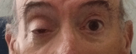
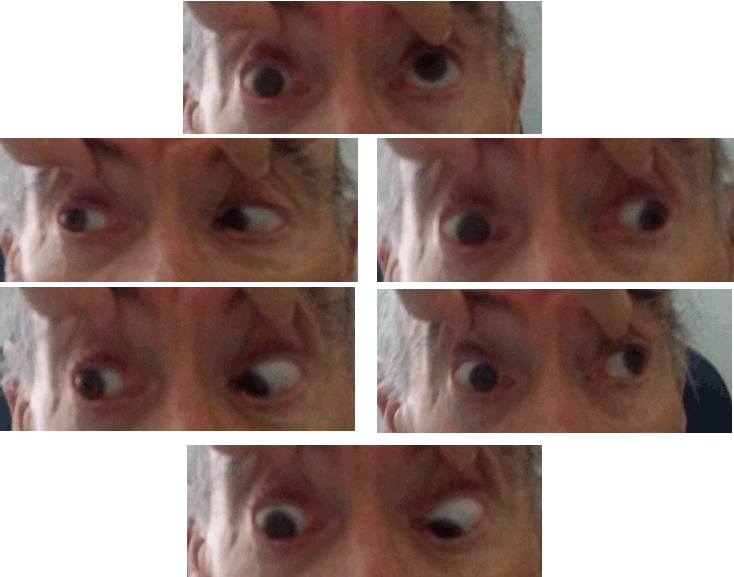
We present the clinical case of a 72-year-old Caucasian male with ophthalmological history of right eye (oculus dexter) amblyopia caused by an untreated esotropia. No relevant medical history was known, with irregular surveillance by his general doctor and no regular medication. The patient presented with holocranial headache, right eye ptosis and temporary diplopia seven days prior to emergency room observation. He denied ocular pain, fever, previous trauma or other systemic or neurologic symptoms. Ophthalmic exam disclosed right eye best corrected visual acuity of the right eye of 20/200 and of the left eye of 20/25. A right ptosis (Figure 1) with attainment of the visual axis, without any inflammatory signs. The pupils were symmetric with a present but sluggish right direct reflex, and a doubtful Marcus Gunn pupil. The patient was orthotropic (Figure 1). The ocular motility examination showed a limitation in adduction, elevation and depression of the right eye (Figure 2). No diplopia was present during observation neither other neurological defects. The slit lamp examination revealed a cataract and funduscopic examination was unremarkable. Computed tomography (CT) scan of the brain and orbits did not reveal acute changes and a analytical study revealed raised inflammatory parameters with neutrophilic leukocytosis (2000, 92.9) and protein chain reaction PCR slightly elevated (4.69). CT angiography that did not reveal aneurysmatic dilations. Brain magnetic resonance imaging (MRI) scan showed countless small foci of enhancement leptomeningeals with cerebellar predominance and additional cerebral foci and in the the internal acoustic canal on T1 acquisition. These findings suggest a possible meningeal carcinomatosis. Lumbar puncture revealed atypical cells (69 mm3), cerebrospinal fluid (CSF) cytology showed neoplastic cells suggesting infiltration by carcinoma (CK 18+). Cerebrospinal fluid serology was negative for Cryptococcus, Lyme disease and syphilis. Cerebrospinal fluid bacteriology was negative. The patient underwent investigation for primary tumor site with CT scan of the neck, chest, abdomen and pelvis. The examination showed multiple lymph nodes on level III and IV referred mainly because of their number and size, the larger one with approximately 21x8.5 mm, and a mass with starting point on the left lateral wall of the bladder, with calcifications, measuring approximately 4x2 cm. The patient was submitted to a flexible cystoscopy that showed a sessil lesion with 3 cm and with probable invasion of the meatus; for a best description a transurethral resection (TUR) of the bladder was proposed. Once made, the histology of the biopsies of TUR revealed fragments of a infiltrative urothelial carcinoma of high grade. The patient started treatment with corticosteroids and methotrexate. The final diagnosis is a palsy of the oculomotor nerve by meningeal carcinomatosis in the context of urothelial carcinoma. The patient was hospitalized in the oncology department and started a systemic treatment, unfortunately with a poor prognosis. | ||||||
| ||||||
| ||||||
|
DISCUSSION
| ||||||
|
The oculomotor nerve supplies somatic: superior rectus, inferior rectus, inferior oblique, medial rectus and levator palpebrae superioris as well as autonomic (papillary sphincter and ciliary) muscles of the eye. Oculomotor nerve palsy may be congenital or acquired, complete or partial, pupil sparing or pupil involving, isolated or accompanied by neurological signs. Precise knowledge of its origin and course from nuclear level to terminal muscles is essential to localize the site of involvement [2]. Third cranial nerve palsy may be the first manifestation of a serious systemic disease and accounts for about one third of presenting cranial nerve palsies. The pupil involvement is commonly related to compressive lesions, in particular aneurysms, on the other hand, pupil sparing third nerve palsy suggest microvascular etiologies [3]. This case reveals an atypical presentation in a patient with previous esotropia and amblyopia and therefore diplopia was not an important symptom for the patient as well as exotropia was not present. For this reason, the patient was orthotropic. The pupil involvement was not obvious so we could consider it as an incomplete palsy (anisocoria was not present), but since there was a sluggish right direct reflex, and a doubtful Marcus Gunn pupil, neuroimaging was mandatory to exclude life-threatening compressive causes. The management of acute partial pupil-sparing is controversial, some suggest that the incidence of aneurysms in this group is low enough that imaging is not necessary, others advocate angiography for all and others suggest a period of observation, since a pupil-sparing partial third nerve palsy from an aneurysm usually develops papillary involvement within a few days [4]. Once excluded aneurysmatic causes further investigation was needed. In this case, the oculomotor nerve palsy appears as a direct compression or infiltration of the oculomotor nerve in his pathway. There are some reports in literature of isolated oculomotor nerve palsy as a paraneoplastic manifestation: gastric diffuse large B cell lymphoma with neoplastic infiltration, although isolated neuropathy is rare, non-small cell lung cancer and Burkitt lymphoma. The majority of the patients with paraneoplastic manifestations and oculomotor nerve palsy exhibited cavernous sinus involvement rather than oculomotor nerve infiltration [5][6][7]. Literature has described a case of a multiple cranial palsies associated with gallbladder cancer [8]. Brain MRI scan combined with cerebrospinal fluid cytology examination is considered optimal for evaluating the cause of oculomotor nerve palsy, but may not be diagnostic in every case. | ||||||
|
CONCLUSION
| ||||||
|
In conclusion, this case demonstrates that isolated oculomotor nerve palsy demands a careful assessment and complete investigation. This case of isolated nuclear oculomotor nerve palsy with atypical features may mimic oculomotor ischemic nerve palsy usually associated with diabetes mellitus and hypertension. The outcome of oculomotor nerve palsy is related to its cause. | ||||||
|
REFERENCES
| ||||||
| ||||||

|
[HTML Abstract]
[PDF Full Text]
|
|
Author Contributions
Filipa Caiado Sousa – Substantial contributions to conception and design, Acquisition of data, Analysis and interpretation of data, Drafting the article, Revising it critically for important intellectual content, Final approval of the version to be published André Diogo Barata – Substantial contributions to conception and design, Drafting the article, Revising it critically for important intellectual content, Final approval of the version to be published Filipa Teixeira – Substantial contributions to conception and design, Drafting the article, Revising it critically for important intellectual content, Final approval of the version to be published Vítor Silva – Analysis and interpretation of data, Revising it critically for important intellectual content, Final approval of the version to be published |
|
Guarantor
The corresponding author is the guarantor of submission. |
|
Source of support
None |
|
Conflict of interest
Authors declare no conflict of interest. |
|
Copyright
© 2017 Filipa Caiado Sousa et al. This article is distributed under the terms of Creative Commons Attribution License which permits unrestricted use, distribution and reproduction in any medium provided the original author(s) and original publisher are properly credited. Please see the copyright policy on the journal website for more information. |
|
|