
|
 |
|
Case Report
| ||||||
| Tubercular thyroid abscess: A case report | ||||||
| Bhavinder Arora | ||||||
|
1Professor, Department of Surgery, Pt B D Sharma University of Health Sciences, PGIMS, Rohtak
| ||||||
| ||||||
|
[HTML Abstract]
[PDF Full Text]
[Print This Article] [Similar article in Pumed] [Similar article in Google Scholar] 
|


| How to cite this article |
| Arora B. Tubercular thyroid abscess: A case report. Int J Case Rep Images 2017;8(9):587–591. |
|
ABSTRACT
| ||||||
|
Introduction:
Thyroid gland tuberculosis is a very rare extrapulmonary presentation of tuberculosis even in countries where pulmonary tuberculosis is endemic. Thyroid gland tuberculosis was reported in 19th century. Thyroid gland tuberculosis presenting as abscess is very rare with occasional case reports in literature. A case of thyroid gland abscess is presented here. Keywords: Abscess, Antitubercular therapy, Fine needle aspiration, Thyroid, Tuberculosis | ||||||
|
INTRODUCTION
| ||||||
|
Tuberculosis of thyroid is a rare manifestation of extra pulmonary tuberculosis. The incidence is very low even in countries where pulmonary tuberculosis is endemic [1]. The incidence of this disease varies from 0.1–1.15% [1][2]. This low incidence of thyroid tuberculosis is attributed high vascularity of thyroid gland and its ability to resist infection [2]. The thyroid gland gets infected by hematogenous and lymphogenous route or direct spread from tubercular cervical lymph nodes [3]. The clinical presentation of tubercular infection of thyroid can be chronic or cold abscess, subacute thyroiditis and rarely as acute abscess. However, the most common manifestation is cold or caseous abscess and a solitary thyroid nodule. Tubercular thyroid can be confused malignant thyroid [4]. The accurate diagnosis needs help of radiological investigations besides elaborate clinical history and examination [5]. Tissue diagnosis either by fine needle aspiration or histopathology is essential [6]. Tubercular thyroid abscess are occasionally available in medical literature. One such case report of tubercular abscess has been presented here. | ||||||
|
CASE REPORT
| ||||||
|
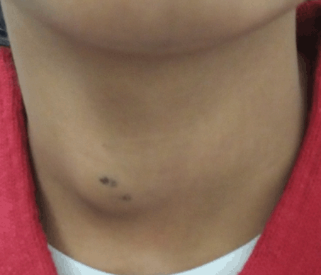
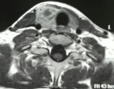
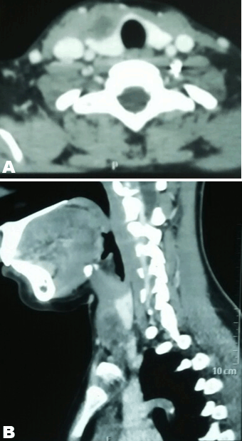
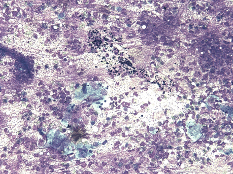
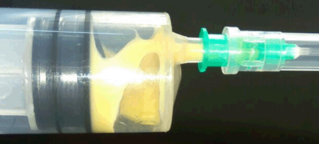
A 16-year-old girl presented with swelling in the right lobe of thyroid. There were no generalized symptoms like fever, malaise, night sweats and weight loss. There was no history of difficulty in deglutition or voice change. On clinical examination there was a single abscess of size 4x4 cm in right lobe of thyroid moving with deglutition. The margins were well demarcated, smooth surface, non-tender and overlying skin was normal with a tattoo mark on it (Figure 1). There were no clinical features of hypothyroidism or hyperthyroidism. Routine blood investigations were done; hemoglobin 11.0 g/dl, total leucocytes count 8600/mm3, neutrophils 67/mm3, lymphocytes 31/mm3 and eosinophils 2/mm3. The erythrocyte sedimentation rate was 20 mm. The Mountax test was highly positive more than 10 mm in diameter. Thyroid function tests T3, T4, TSH were normal. X-ray chest was normal. Ultrasonography of neck revealed a 50x45x20 mm solitary nodule in the right lobe of liver. This solitary thyroid nodule was showing thick irregular wall with central necrosis reported as suspected thyroid abscess. Magnetic resonance imaging scan of the neck showed a lesion of intermediate signal intensity due to presence of dense inflammatory cells and granulomas with central necrosis (Figure 2). A doubt about carcinoma of thyroid was placed as differential diagnosis. The CECT scan of neck was done to rule out carcinoma of thyroid gland. This was helpful in diagnosis of tubercular thyroid abscess as localized caseous lesion in right lobe of thyroid (Figure 3). Fine needle aspiration from this solitary thyroid nodule was done to confirm the diagnosis. The stained smears revealed degenerated and intact neutrophils, and macrophages in serofibrinous background. A few epithelioid granuloma and multinucleated giant cells are also seen suggestive of tuberculosis with central caseous necrosis. Ziehl–Neelsen staining with 20% H2SO4 was noncontributory (Figure 4). From the central part of swelling about 2 ml of thick yellow color pus was aspirated as shown in Figure 5. The smears prepared from this pus did not show any acid-fast bacilli. After aspiration the swelling decreased in size. The cytological diagnosis of tubercular abscess was made. The patient was put on antitubercular treatment with four drug regimens. The swelling decreased in size in next three months (Figure 6). She was asked to continue on three drug regimens for another six months leading to complete resolution of swelling. | ||||||
| ||||||
| ||||||
| ||||||
| ||||||
| ||||||
| ||||||
|
DISCUSSION
| ||||||
|
Tuberculosis of thyroid is rare diagnosis reported since early 19th century [7]. It is a rare extrapulmonary manifestation of tuberculosis, the true incidence of tuberculosis of thyroid is unknown. This rare involvement of thyroid gland to tubercular infection is attributed to high vascularity of thyroid gland and bactericidal property of colloid material [8]. Thyroid tuberculosis may be primary or secondary concurrent with pulmonary tuberculosis. The primary involvement of thyroid without pulmonary involvement is extremely rare [9]. There are two routes of infection by which thyroid gland get infected; generalized dissemination by hematogenous route as in miliary tuberculosis and a focal spread to thyroid gland. Focal spread may be primary of thyroid gland called primary tuberculosis of thyroid gland [10]. However, it may be secondary from adjacent lymph node [11]. Occasionally, there may be lymphogenous spread. The clinical presentation of tubercular thyroid abscess is generally as a solitary thyroid nodule or a cold abscess rarely presenting as acute abscess [12][13]. Solid thyroid nodule can mimic clinically as thyroid carcinoma [14]. Presenting symptoms in tubercular thyroid abscess are variable. Most of the patients present as solitary thyroid nodule with no sign of acute inflammation. All main diseases of thyroid gland should be considered including carcinoma thyroid [15]. The accurate diagnosis of tubercular thyroid has to be made by using investigation. The ultrasound is the basic investigation for a solitary thyroid nodule. Radiological imaging techniques of MRI scan and CT scan make the diagnosis of thyroid tuberculosis and also rule out malignancy of thyroid [16]. However, confirmation of diagnosis can only be done using tissue diagnostic technique of fine needle aspiration cytology [17]. The cytological diagnosis can be made by presence of tubercular granuloma. Acid-fast bacilli staining may not detect tubercular bacilli [18]. In such cases PCR can be done [19]. The confirmatory diagnosis of tuberculosis of thyroid by fine needle aspiration cytology can avoid unnecessary thyroid surgery for histopathological confirmation. Antitubercular therapy is the preferred method of treatment of tuberculosis of thyroid nowadays [20]. If thick pus is present in central part it can be aspirated using thick needle. Repeated aspiration may be necessary. A few cases in which pus cannot be aspirated open drainage may be necessary. Repeated needle drainage and antitubercular drug therapy is the treatment of choice as being the least invasive method [21]. However, very large thyroid abscess may need open drainage and excision is required rarely. Those patients who do not respond to antitubercular therapy in three months duration may need surgical excision of thyroid nodule or hemithyroidectomy [22]. | ||||||
|
CONCLUSION
| ||||||
|
Tubercular thyroid abscess is a rare clinical diagnosis as pulmonary tuberculosis may not be associated with pulmonary tuberculosis in most of these patients. Tubercular thyroid abscess can be diagnosed only with a very high degree of clinical suspicion. Imaging techniques like magnetic resonance imaging scan and computed tomography scan are useful in making the diagnosis of tubercular thyroid abscess. Definitive diagnosis can be made by cytological examination by presence of tubercular granuloma. Tubercular thyroid abscess can be treated by aspiration of pus followed by antitubercular treatment thus avoiding surgery of thyroid. | ||||||
|
REFERENCES
| ||||||
| ||||||


|
[HTML Abstract]
[PDF Full Text]
|
|
Author Contributions
Bhavinder Arora – Substantial contributions to conception and design, Acquisition of data, Analysis and interpretation of data, Drafting the article, Revising it critically for important intellectual content, Final approval of the version to be published |
|
Guarantor
The corresponding author is the guarantor of submission. |
|
Source of support
None |
|
Conflict of interest
Authors declare no conflict of interest. |
|
Copyright
© 2017 Bhavinder Arora. This article is distributed under the terms of Creative Commons Attribution License which permits unrestricted use, distribution and reproduction in any medium provided the original author(s) and original publisher are properly credited. Please see the copyright policy on the journal website for more information. |
|
|