
|
 |
|
Case Report
| ||||||
| Early laparoscopic treatment of an obstructed paracecal hernia in an octogenarian: A case report | ||||||
| Robert Cooke1, Nicholas Heywood1, Abhiram Sharma1, Velauthan Rudralingham1 | ||||||
|
1University Hospital of South Manchester, Southmoor Road, Wythenshawe, Manchester
| ||||||
| ||||||
|
[HTML Abstract]
[PDF Full Text]
[Print This Article] [Similar article in Pumed] [Similar article in Google Scholar] 
|


| How to cite this article |
| Cooke R, Heywood N, Sharma A, Rudralingham V. Early laparoscopic treatment of an obstructed paracecal hernia in an octogenarian: A case report. Int J Case Rep Images 2017;8(9):571–574. |
|
ABSTRACT
| ||||||
|
Paracecal hernias are a rare cause of small bowel obstruction. The management of these has been predominantly by laparotomy. An 84-year-old male was presented who underwent early laparoscopic management of a paracecal hernia. This resulted in avoidance of laparotomy and a short postoperative hospital stay. Keywords: Paracecal hernia, Bowel obstruction, Laparoscopy | ||||||
|
INTRODUCTION
| ||||||
|
Small bowel obstruction is a common presentation to the acute surgical take. It is most commonly due to adhesions, however up to 5.8% of cases are caused by internal hernias. Of which paracecal hernias account for up to 6.6% [1][2]. Clinical diagnosis can be difficult especially in the virgin abdomen as signs are non-specific. Computed tomography scan is the current main tool for diagnosis but the exact findings are only usually identified during surgery. Laparotomy has been the mainstay for management for these patients [3]. | ||||||
|
CASE REPORT
| ||||||
|
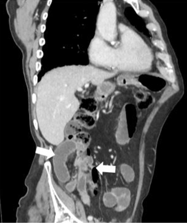
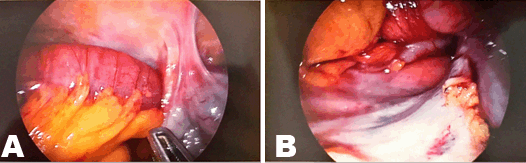
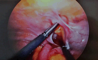
An 84-year-old male with no previous history of abdominal surgery presented to accident and emergency department with a one day history of intermittent abdominal pain. He denied any nausea or vomiting, however, noted a reduced stool frequency in the preceding two weeks. His past medical history included ischemic heart disease, cerebrovascular disease, diabetes, hypertension, pulmonary fibrosis, benign prostatic hyperplasia and he had an abdominal aortic aneurysm under surveillance. Initial assessment of the patient’s abdomen revealed no tenderness or distension and physiological observations were within normal limits. Blood results were unremarkable except for a mildly elevated C-reactive protein of 42. During a period of observation he experienced increasing pain and developed tenderness in the right iliac fossa. A computed tomography scan was performed, revealing a localized incarcerated segment of small bowel lateral to the cecum in the right paracolic gutter with upstream dilatation of small bowel loops lying anterior to the ascending colon. The associated localized mesenteric congestion and fluid surrounding the localized segment of small bowel in the right iliac fossa were suggestive of a paracecal hernia. The patient was taken to theatre for laparoscopy within 24 hours of admission. A paracecal hernia containing an obstructing loop of jejunum was identified between the cecum and lateral abdominal wall. Multiple adhesions in this area created a narrow necked blind ending ‘hernia cave’ in the right paracolic gutter. An adjacent internal hernia contained an unobstructed vermiform appendix. Laparoscopic reduction of the small bowel from the hernia was performed and adhesiolysis undertaken to prevent recurrence. The patient had an uncomplicated postoperative course, returned to baseline function and was discharged on the third postoperative day. | ||||||
| ||||||
| ||||||
| ||||||
| ||||||
|
DISCUSSION
| ||||||
|
A hernia is defined as the protrusion of a viscus through a defect in the wall of the cavity in which it resides. When bowel passes through a peritoneal defect or mesenteric aperture, it is known as an internal hernia [4]. Internal hernias account for up to 5.8% of presentations of small bowel obstruction, of which paracecal hernias make up only 6.6% [1]. The paracecal peritoneum develops once the midgut has completed migration. The result is formation of four paracecal recesses being formed. These are the superior ileocecal recess, inferior ileocecal recess, retrocecal recess and paracolic sulci. These all have the potential to form hernial orifices [5]. Symptoms of abdominal pain and vomiting from paracecal hernias are variable and depend on the degree of incarceration or obstruction. Often due to their anatomical location, they may be mistaken for other right iliac fossa pathology, such as appendicitis [1][4]. When there is diagnostic uncertainty, or if internal hernia is suspected, computed tomography scan is the preferred investigation of choice. Aside from confirming the diagnosis, this readily available imaging provides information about the presence of strangulation and ischemia as an aid to preoperative planning. The presence of a cluster of dilated small bowel loops, a transition point lateral to the cecum and congestion of mesenteric vessels raise suspicion for an obstructing paracecal hernia. The cecum can sometimes be seen to have been displaced anteromedially. Other imaging modalities such as plain X-ray films and contrast studies often provide limited information and may result in delayed diagnosis subsequently increasing the risk of complications [2][4][6] Laparotomy has generally been the preferred approach for the presence of small bowel obstruction, however, many studies including O’Connor et al. review of over 2000 cases have shown the safety and efficacy of laparoscopic surgery [7]. The debate over the approach is on-going because of the technical difficulties and potential complications associated with laparoscopic surgery in these patients. These include damage to distended small bowel loops and visualization of the transition point, making a high conversion rate to laparotomy and morbidity. However, benefits of laparoscopic surgery in small bowel obstruction have been shown, these are reduction in postoperative hospital stay, reduction in morbidity, reduction in adhesion formation and faster return to bowel function [7]. A recent review of published cases of paracecal hernias by Ogami et al. [3] highlighted that the vast majority of reported cases have been managed by laparotomy and that only three cases have been managed laparoscopically, none of which were performed within the UK. On reviewing these three case reports [3] [8] [9] all were performed after several days of conservative treatment which had not responded, in comparison to our patient who was operated on within 24 hours. All the cases showed viable bowel upon reduction of the hernia and no resections were required. The postoperative courses were uneventful with patients being discharged between 9 and 10 days postoperatively. In our case, the prompt diagnosis and early laparoscopic intervention had a reduced length of stay in comparison to the previously discussed cases. | ||||||
|
CONCLUSION
| ||||||
|
We have demonstrated the safe use of early laparoscopic reduction of paracecal hernia and division of adhesions in an elderly patient. We have avoided the need for laparotomy in this patient resulting in early return to function, avoiding the morbidity associated with open abdominal surgery. Early computed tomography scan and laparoscopic approach should be considered in this group of patients. | ||||||
|
REFERENCES
| ||||||
| ||||||


|
[HTML Abstract]
[PDF Full Text]
|
|
Author Contributions
Robert Cooke – Substantial contributions to conception and design, Acquisition of data, Analysis and interpretation of data, Drafting the article, Revising it critically for important intellectual content, Final approval of the version to be published Nicholas Heywood – Analysis and interpretation of data, Revising it critically for important intellectual content, Final approval of the version to be published Abhiram Sharma – Analysis and interpretation of data, Revising it critically for important intellectual content, Final approval of the version to be published Velauthan Rudralingham – Analysis and interpretation of data, Revising it critically for important intellectual content, Final approval of the version to be published |
|
Guarantor
The corresponding author is the guarantor of submission. |
|
Source of support
None |
|
Conflict of interest
Authors declare no conflict of interest. |
|
Copyright
© 2017 Robert Cooke et al. This article is distributed under the terms of Creative Commons Attribution License which permits unrestricted use, distribution and reproduction in any medium provided the original author(s) and original publisher are properly credited. Please see the copyright policy on the journal website for more information. |
|
|