| |
|
|
|
Case Report
| ||||||
| Necrotizing fasciitis of lower limb: Is it ischemia or E. coli infection? | ||||||
| Takahiro Matsuo2, Hiroki Mochizuki1, Atsushi Mizuno1, Keiichi Furukawa2, Yutaro Nishi1, Koichiro Niwa1 | ||||||
|
1Department of Cardiology, St. Luke's International Hospital, Tokyo, Japan.
2Department of Infectious Diseases, St. Luke's International Hospital, Tokyo, Japan. | ||||||
| ||||||
|
[HTML Abstract]
[PDF Full Text]
[Print This Article]
[Similar article in Pumed] [Similar article in Google Scholar] 
|
| How to cite this article |
| Matsuo T, Mochizuki H, Mizuno A, Furukawa K, Nishi Y, Niwa K. Necrotizing fasciitis of lower limb: Is it ischemia or E. coli infection? Int J Case Rep Images 2016;7(11):701–705. |
|
Abstract
|
|
Introduction:
Necrotizing fasciitis (NF) is a life-threatening soft-tissue infection characterized by a fulminant course and high mortality. Diagnosis of necrotizing fasciitis is often difficult, because nascent necrotizing fasciitis often appears deceptively benign and lack specific diagnostic clues. In addition, the manifestations of NF are similar to those of acute limb ischemia. Here, we introduce the case of necrotizing fasciitis accompanied with acute limb ischemia on left lower leg.
Case Report: A 71-year-old male with a history of stomach cancer complained of vomiting and dyspnea one day before admission to our hospital. Two days after admission, his left lower extremity whitened and became edematous. Contrast-enhanced computed tomography revealed severe stenosis of the left common iliac artery. We were unable to rule out acute limb ischemia and performed endovascular revascularization. However, the leg did not improve and major amputation was performed. The leg pathology was suggestive of necrotizing fasciitis. Conclusion: We should not forget the possibility that any systemic infection can result in necrotizing fasciitis even if the patients have high risk of acute limb ischemia. | |
|
Keywords:
Acute limb ischemia, E. coli, Escherichia coli, Necrotizing fasciitis, Pneumonia
| |
|
Introduction
| ||||||
|
Necrotizing fasciitis (NF) is infrequent but highly lethal infection and is associated with systemic toxicity and a mortality rate of 30–60% [1] [2]. It affects the superficial fascia and subcutaneous tissue. Establishing the diagnosis of NF is difficult because nascent NF often appears deceptively benign and lack specific diagnostic clues [3]. Delay of diagnosis leads to delayed surgical debridement, which leads to higher mortality [1]. The manifestations of NF are similar to those of acute limb ischemia. Here, we introduce the case of necrotizing fasciitis accompanied with acute limb ischemia on left lower leg. | ||||||
|
Case Report
| ||||||
|
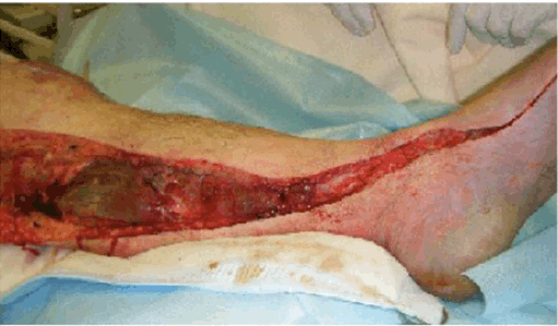
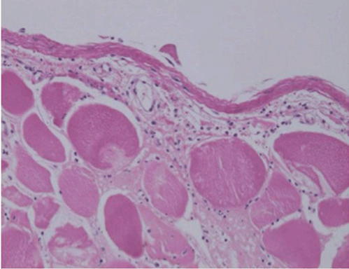
A 71-year-old male with a history of hypertension and stomach cancer complained of vomiting and dyspnea one day before admission to hospital. On admission, he was unconscious, with a Glasgow coma score of 3. Clinical examination revealed that the patient's state was critical, with a temperature of 39.3ºC, blood pressure 61/39, heart rate 144/min, respiratory rate 43/min, and oxygen saturation 94% on a non-rebreather mask at 15 L/min oxygen. The patient's left-sided respiratory sounds were decreased, with coarse crackles. The bowel sounds were normal and the abdomen was soft and non-tender. We intubated him in the emergency room because the patient was in shock with impaired consciousness. Laboratory findings revealed an elevated white blood cell (WBC) count of 1200/µL, C-reactive protein 27.1 mg/dL, creatinine kinase (CK) 2405 U/L, and creatinine 1.62 mg/dL. Arterial blood gas analysis revealed significant lactic acidosis (Table 1). Computed tomography (CT) scan revealed consolidation in the superior lobe of the left lung. Immunochromatographic membrane tests using urine sample for rapid detection of Streptococcus pneumonia antigen and Legionella antigen (BinaxNOW S. pneumoniae and BinaxNOW Legionella; Inverness Medical Innovations), were performed but gave negative results. Gram staining of the sputum revealed Gram-negative rods with neutrophils. The patient was diagnosed with pulmonary insufficiency associated with pneumonia and septic shock and admitted to the intensive care unit (Figure 1). We initiated therapy with vasopressors and broad-spectrum antibiotics (piperacillin/tazobactam and ciprofloxacin). On the same day, the patient's blood culture became positive for bacteremia with a Gram-negative rod. Therefore, direct hemoperfusion with a polymyxin B–immobilized fiber column was initiated. On day-4, the skin of the left lower leg abruptly turned white and purpura developed. Doppler ultrasound was unable to detect blood flow in the left dorsalis pedis artery, and a CT scan showed severe stenosis of the left common iliac and femoral arteries. The patient's CK level had risen to 60,000 IU/L. The clinical manifestations, laboratory findings, and results of radiological imaging were suggestive of acute limb ischemia. We performed angiography, which revealed significant stenosis of the left common iliac artery (Figure 2); this finding was compatible with acute limb ischemia. We discussed whether or not we should perform revascularization because we did not know the precise time of onset of the limb ischemia. We reached the decision to insert a self-expanding stent into left common iliac artery and to perform a counter-incision (Figure 3) because the intracompartmental pressure of the left lower extremity was 70 mmHg. The patient's scrotum had also become swollen and erythematous on day-4, and its color had changed to purple on day-5 (Figure 4). We suspected Fournier gangrene and consulted urologists accordingly. Puncture of the scrotum revealed a serous, yellow, clear fluid, which was compatible with hydrocele testis, not Fournier gangrene. Despite the counter-incision, eight days after the patient's admission the color of his left lower extremity had not improved and the CK had risen rapidly further to 1, 500, 000 U/L. We considered other differential diagnosis including purpura fulinans caused by Streptococcus pneumonia, Streptococcus pyogenes, Vibrio vulnificus, and Neisseria meningitides. We therefore performed major amputation above the hip joint. The patient's general condition, including his vital signs, disseminated intravascular coagulation score, and oxygenation, improved temporarily after this intervention. Unfortunately, two weeks later the patient developed candidemia as a complication of infection from his central venous catheter. Despite the administration of liposomal amphotericin-B to treat the candidemia, the patient died on hospitalization day-38 from multiple organ failure. Pathology | ||||||
| ||||||
|
| ||||||
| ||||||
|
| ||||||
| ||||||
| ||||||
|
Discussion
| ||||||
|
This patient developed lower extremity NF accompanied by severe sepsis and acute pneumonia caused by E. coli. Diagnosis of NF was difficult in this case, because the leg manifestations were similar to those of acute limb ischemia. Pathological examination of the leg finally helped us to differentiate NF from mere limb ischemia. Diagnosing necrotizing fasciitis Although macroscopic findings such as skin discoloration, blisters or bullae, crepitus upon palpation, skin necrosis, and subcutaneous gas [3] [4] are considered typical signs of NF, these findings are similar to those in acute limb ischemia [5]. Microscopic findings such as the presence of WBCs around the fascia and fatty tissue are helpful in establishing a diagnosis of NF, but it takes several days to get the results of such examinations. Indeed, in our case, it took several days to confirm NF in the specimen obtained during surgery. Imaging modalities such as ultrasonography, CT, and magnetic resonance imaging are also considered useful. These imaging studies have revealed that increasing thickness of the fascial layer with or without enhancement is a typical sign but is not specific to the diagnosis of NF, especially when a differential diagnosis of acute limb ischemia is being considered [6]. Among laboratory findings, the laboratory risk indicator for necrotizing fasciitis (LRINEC) score, which includes C-reactive protein, WBC count, hemoglobin level, sodium level, and glucose level, has been developed to distinguish NF from non-necrotizing soft tissue infections, but this scoring system is not helpful in differentiating NF from acute limb ischemia [7]. There are three categories of LRINEC score (points, probability of NF): low (<5, <50%), intermediate (6–7, 50–75%), and high (>8, >75). Although this score is not helpful in differentiating acute limb ischemia, it might have been useful as a reminder of the potential diagnosis of NF early on in this case. It is important not to miss the diagnosis of NF in every high-risk patient, even if acute limb ischemia is suspected, because the macroscopic findings are similar. Necrotizing fasciitis associated with E. coli pneumonia and E. coli bacteremia In this case, the unusual, undetermined origin of the E. coli bacteremia associated with the patient's shock status, together with the pneumonia revealed by the CT imaging, misled us in such a way that we did not diagnose NF early. Early diagnosis and surgical intervention are needed to treat NF patients. We should not forget the possibility that any systemic infection can result in NF, and we should use caution in our clinical approach to skin color changes similar to those found in acute limb ischemia. | ||||||
|
Conclusion
| ||||||
|
Although the diagnosis of necrotizing fasciitis (NF) is often difficult, the use of three tools-macroscopic/microscopic tools, imaging studies, and laboratory findings-can help with the diagnosis. It is important not to miss the diagnosis of NF in every high-risk patient, even if acute limb ischemia is suspected. In addition, considering systemic infection is also important when sputum culture grew E. coli. | ||||||
|
References
| ||||||
| ||||||
|
[HTML Abstract]
[PDF Full Text]
|
|
Author Contributions
Takahiro Matsuo – Substantial contributions to conception and design, Acquisition of data, Analysis and interpretation of data, Drafting the article, Revising it critically for important intellectual content, Final approval of the version to be published Hiroki Mochizuki – Analysis and interpretation of data, Revising it critically for important intellectual content, Final approval of the version to be published Atsushi Mizuno – Analysis and interpretation of data, Revising it critically for important intellectual content, Final approval of the version to be published Keiichi Furukawa – Analysis and interpretation of data, Revising it critically for important intellectual content, Final approval of the version to be published Yutaro Nishi – Analysis and interpretation of data, Revising it critically for important intellectual content, Final approval of the version to be published Koichiro Niwa – Analysis and interpretation of data, Revising it critically for important intellectual content, Final approval of the version to be published |
|
Guarantor of submission
The corresponding author is the guarantor of submission. |
|
Source of support
None |
|
Conflict of interest
Authors declare no conflict of interest. |
|
Copyright
© 2016 Takahiro Matsuo et al. This article is distributed under the terms of Creative Commons Attribution License which permits unrestricted use, distribution and reproduction in any medium provided the original author(s) and original publisher are properly credited. Please see the copyright policy on the journal website for more information. |
|
|