| |
|
|
|
Letters to the Editor
| ||||||
| Long-term outcome in a patient with severe postoperative osteomyelitis of the critical distal third of lower leg | ||||||
| Ingo Schmidt | ||||||
|
SRH Poliklinik Gera GmbH, Straße des Friedens 122, 07548 Gera, Germany
| ||||||
| ||||||
|
[HTML Abstract]
[PDF Full Text]
[Print This Article]
[Similar article in Pumed] [Similar article in Google Scholar] 
|
| How to cite this article |
| Schmidt I. Long-term outcome in a patient with severe postoperative osteomyelitis of the critical distal third of lower leg. Int J Case Rep Images 2016;7(10):680–682. |
|
To the Editors,
|
|
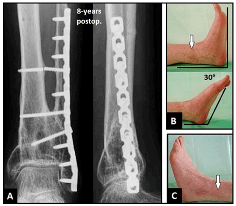
Postoperative osteomyelitis (POM) following highly comminuted fractures of the distal third of lower leg represents a challenging problem. A 56-year-old female presented with an extraarticular closed fracture of the left leg that was treated initially with open reduction and internal fixation (ORIF) (Figure 1A). The patient developed severe POM (Staphylococcus aureus) with soft tissue defect of the distal tibia that was primary treated with removal of all internal osteosynthesis plates, meticulous bony debridement, incorporation of polymethyl methacrylate (PMMA) beads containing gentamycin, multiple negative-pressure vacuum assisted closure (VAC) therapies, and external fixation (Figure 1B). After microbiological consolidation of POM, assessed by culture and histology, the soft tissue defect was covered by a distally based sural flap. After wound healing (Figure 1C), the PMMA beads and external fixation were removed combined with filling of the cavity using cancellous iliac crest bone grafts (CICBG), and ORIF of the distal fibula combined with a distal tibiofibular fusion (DTFF) using CICBG and three screws was performed (Figure 1D). In the further course, the patient developed aseptic soft tissue necrosis with exposure of osteosynthesis plate of the distal fibula that was covered by a distally based peroneus brevis muscle flap combined with split skin grafts. Three months after injury, including 18 surgical procedures, the patient could be mobilized with full weight-bearing on the affected leg (Figure 1E). Follow-up on eighth year, the DTFF showed unchanged complete union in the absence of varus or valgus deformity as well as post-traumatic arthritis in the ankle, note that the proximal screw for DTFF was broken without needing of removal (Figure 2A). The dorsal-plantar motion arc in the ankle was 30°, and both flaps were healed without any complications (Figure 2B-C). Reconstruction of the distal third of lower leg is associated with the highest complication rate for local surgical procedures. The three-stage surgical treatment for POM of this site, that includes meticulous bony debridement/soft tissue coverage/bony reconstruction with additional antibiotic therapy, has proven to be effective [1]. Negative-pressure VAC therapy before soft tissue coverage provides a sterile and controlled environment that can lessen the duration of wound healing, promotes better capillary circulation, and decreases the bacterial load [2]. To avoid posttraumatic arthritis in the ankle, DTFF has proven to be as one suitable and reliable option for its stabilization [3]. The use of distally based sural and peroneus brevis muscle flaps for coverage of the distal end of lower leg is recommended for small soft tissue defects and/or exposure of bones in patients who are not willing or healthy enough to undergo free microvascular tissue transplantation, and do not require microsurgical expertise [4] [5]. Keywords: Distal third lower leg, Postoperative osteomyelitis, Distally based sural flap, Distal tibiofibular fusion, Distally based peroneus brevis muscle flap |
|
|
|
|
|
|
|
References
|
|
|
[HTML Abstract]
[PDF Full Text]
|
|
Author Contributions:
Ingo Schmidt – Substantial contributions to conception and design, Acquisition of data, Analysis and interpretation of data, Drafting the article, Revising it critically for important intellectual content, Final approval of the version to be published |
|
Guarantor of submission
The corresponding author is the guarantor of submission. |
|
Source of support
None |
|
Conflict of interest
Authors declare no conflict of interest. |
|
Copyright
© 2016 Ingo Schmidt. This article is distributed under the terms of Creative Commons Attribution License which permits unrestricted use, distribution and reproduction in any medium provided the original author(s) and original publisher are properly credited. Please see the copyright policy on the journal website for more information. |
|
|