| |
|
|
|
Case Report
| ||||||
| An unusual presentation of odontogenic keratocyst with malignant changes in a 13-year-old girl | ||||||
| Manal Ibrahim Elnouaem1, Zeinab El Sayed Darwish2, Rana Aly Sharaf3 | ||||||
|
1Professor Oral Pathology, Phd, Professor, Oral Pathology Department, Faculty of Dentistry, Alexandria University, Alexandria, Egypt.
2Professor Oral Pathology, Phd, Oral Pathology Department, Faculty of Dentistry, Alexandria University, Alexandria, Egypt. 3Demonstrator, Oral medicine and Diagnosis, Demonstrator, Oral medicine and diagnosis Department, Faculty of Dentistry, Alexandria, University, Alexandria, Egypt. | ||||||
| ||||||
|
[HTML Abstract]
[PDF Full Text]
[Print This Article]
[Similar article in Pumed] [Similar article in Google Scholar] 
|
| How to cite this article |
| Elnouaem MI, El Sayed Darwish Z, Aly Sharaf R. An unusual presentation of odontogenic keratocyst with malignant changes in a 13-year-old girl. Int J Case Rep Images 2016;7(10):657–661. |
|
Abstract
|
|
Introduction:
Odontogenic keratocysts as defined by WHO are known for their characteristic behavior, high tendency to recur, unusual pattern of growth and different treatment methods. Squamous cell carcinoma on top of keratocysts is extremely rare and is mostly limited to jaw bones.
Case Report: We hereby report, not previously reported a characteristic case of an odontogenic keratocyst related to the anterior maxillary teeth in a teenage female of 13 years. On histological examination of the biopsy, frank invasion was seen and a diagnosis of squamous cell carcinoma on top of keratocyst was given. Conclusion: The diagnosis was predicted on recognition of the histopathologic features of odontogenic keratocyst associated with dysplastic features of the epithelial lining and areas of invasion in other parts; the clinical symptoms are modest, only presenting a palatal swelling. | |
|
Keywords:
Children population, Jaw bones, Odontogenic keratocyst, Squamous cell carcinoma
| |
|
Introduction
|
|
Cysts of the jaws tend to occur across different age and ethnic groups. Scarce information on those which occur in the pediatric population had been reported in literature. In some studies, pediatric jaw cysts showed predominance of developmental cysts above inflammatory ones [1] or have a predilection for inflammatory cyst occurrence above developmental ones [2]. Odontogenic keratocysts (OKC) have been first introduced in 1953 by Philipsen. They comprise 3–10.5% of all jaw cysts occurring mainly in the 2nd and 3rd decades of life. Majority of keratocysts occur in the mandible but may occur in any part of the jaw. They are solitary unless they are a part of nevoid basal cell carcinoma syndrome [3]. Malignant transformation of odontogenic keratocysts has frequently been associated with a painless benign expansile lesion of the jaws presenting a radiographic picture similar to cystic lesions from which they arise [4]. This case report is about a 13-year-old girl with a keratocyst which showed malignant transformation. |
|
Case Report
|
|
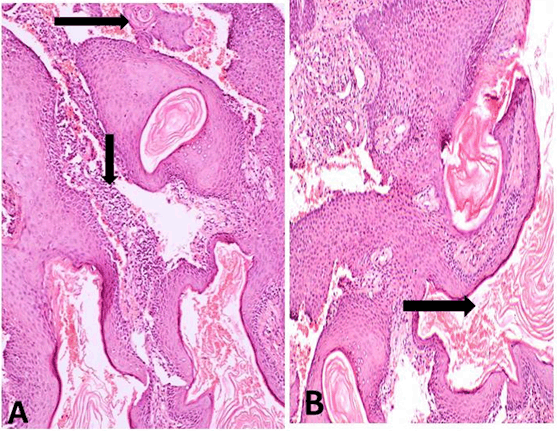
A 13-year-old girl patient was reported to our dental college complaining of a symptomless progressive enlargement of the palatal mucosa accompanied by an abnormal taste for last six months. Clinical intraoral examination revealed a bony swelling of 1.5x2 cm mainly bulging buccally related to the right maxillary anterior teeth. The overlying mucosa was reddish with a minor degree of laceration. On palpation the swollen area showed egg shell crackling. Aspiration of the swelling yielded turbid cystic fluid. Radiographic examination based on the available panoramic view revealed a unilocular expansile lucency which was well defined and partially corticated. The radiolucency involved the roots of both maxillary central and lateral incisors and extended up to premolar area. No destructive osseous changes were, seen however, mild resorption of the roots of #11 and #12 was noted; along with a marked divergence of the roots of #12 and #14. The radiolucency was related to horizontally impacted tooth (Figure 1). There was no medical history of significance. Based on the clinical and radiographic findings, the initial diagnosis of odontogenic cyst was made and the lesion was subjected to histological examination. Histopathologic examination revealed a uniformly corrugated parakeratinized epithelial lining, however, some other areas showed partially uniform thickness with palisaded basal cells. A huge amount of keratin was seen filling the cystic cavity (Figure 2A-B). The epithelium exhibited basilar hyperplasia, hyperchromatism, pleomorphism, (Figure 2B), intraepithelial pearl formation (Figure 3A-B). There was discontinuity of basement membrane together with loss of polarity of basal cells in some areas (Figure 3B). The underlying stoma showed few epithelial cell nests and keratin pearls. Scattered chronic inflammatory cell infiltrate was present in the fibrous tissue capsule (Figure 4A-B) These histological findings led us to the diagnosis of odontogenic keratocyst which has undergone malignant transformation giving the diagnosis of well differentiated squamous cell carcinoma on top of keratocyst according to the presence of frank point of invasion as well as epithelial and keratin pearls in the underlying stoma. |
|
|
|
|
|
|
|
|
|
Discussion
|
|
Keratocysts are locally destructive lesions which occur intrabony and tend to enlarge to reach a significant size before it appears as a swelling buccolingually thus preventing early diagnosis of any underlying lesion [5]. In this case a 13-year-old girl having an uncommon finding; keratocysts in the pediatric populations are under reported due to the loss of primary teeth thus resulting in resolution of some cystic lesions which are asymptomatic and of limited size especially when not involving the underlying tooth follicles of permanent successors. The exfoliation of primary teeth may limit the development and expansion of these cystic lesions which increases the rarity of reported incidence of developmental and inflammatory cysts in pediatric population when compared to adult population [1]. Multiple odontogenic keratocysts of the jaws are known to be associating other features as vertebral and rib anomalies and intra cranial calcification in basal cell nevus syndrome. In this syndrome, odontogenic keratocysts occur more commonly in the 2nd and 3rd decades of life which is ten years earlier than the more common solitary isolated ones [6]. As it is rare to see a single odontogenic keratocyst in a 13-year-old girl, this raised the suspicion of the presence of the cyst with other associated features of the syndrome, which was excluded by thorough medical examination for vertebral anomalies and nevoid basal cell carcinomas of the skin, epidermal cysts or skin pits. Generally no cardinal signs of syndrome were detected in the present case. Therefore, the basal cell nevus syndrome was ruled out. The present case was located in the anterior region of the maxilla. This is in accordance with the previously reported studies [1] [2]. However, mandibular predominance was reported by others [7]. Neither age nor site of the present case matched the common age or site of odontogenic keratocysts where the common age is 20–40 years and the mandible is four times more common than maxilla, mostly in posterior mandible and ascending ramus [3] [5]. The occurrence of odontogenic keratocyst in anterior maxilla is relatively rare, it is uncommon to see it in a 13-year-old girl as seen in this study. Intraoral examination revealed a painless slight palatal swelling with expansion of buccal cortical plate with egg shell crackling felt on palpation. The overlying mucosa was red and swollen, Aspiration revealed a turbid fluid which was cheesy in nature similar to keratin. Same clinical features were described by Suma et al. [5] who described the same features in their case of odontogenic keratocyst of the maxillary premolar region. The expansion of the odontogenic keratocyst in the present case may indicate a long standing cyst with no early symptoms and may also indicate the malignant changes which had occurred in the cyst lining thus increasing the rate of growth. The histopathological features in the present case showed carcinomatous changes in the lining of the odontogenic keratocyst. Carcinoma arising on top of this odontogenic keratocyst was unexpected, as in other reported cases where the features were also unexpected. No signs of nerve paresthesia or pain were encountered probably because the malignant changes might be confined to the cyst wall and they maybe still early in their development which may explain the well defined radiolucency in the panoramic X-ray [5]; unlike the findings by Falaki et al. [8] who mentioned that malignant changes in odontogenic cysts should be considered if the radiolucent area has jagged or irregular margins with indentations and ill defined borders, a finding which does not go with the present case. The plain radiograph of the cyst is similar to benign cystic lesions which made Cavalcanti et al. [9] stated that CT scan would be more sensitive tool for detecting carcinomatous changes as it would show the destruction of adjacent bone, if present. The present case had only a panoramic radiograph which was taken to detect the lesion and in which features of malignancy were not evident. The odontogenic keratocyst has been one of the most controversial entities of the maxillofacial region since described by Phillipsen. WHO recently classified odontogenic keratocysts as benign neoplasms recommending the term keratocystic odontogenic tumor (KCOT). Rarely documented the epithelial lining of KCOT showed features of dysplasia and malignant transformation [10] which made this case a rare one where malignant changes were present together with the fact the case is in a child. This case report points out to clinical signs, symptoms, radiographic features and histological features of a malignant transformation of an odontogenic keratocyst of anterior maxilla in a 13-year-old child. This case indicates the importance of microscopic examination of all excised cysts. |
|
Conclusion
|
|
This case report analyzed the clinical signs and symptoms, radiographic features and histopathologic appearance of an odontogenic keratocyst in the anterior maxilla of a child which is a rare finding proven that it is not syndrome related. This report illustrates the importance of careful microscopic investigation in all excised cysts because of the possibility of carcinomatous changes despite the relatively benign picture seen in the associating radiograph. |
|
Acknowledgements
|
|
We would like to thank Dr Mervet Moussa; Professor of Oral Pathology, Cairo University for her help and continuous support. |
|
References
|
|
|
Suggested reading
|
|
|
[HTML Abstract]
[PDF Full Text]
|
|
Author Contributions
Manal Ibrahim Elnouaem – Substantial contributions to conception and design, Acquisition of data, Analysis and interpretation of data, Drafting of the article, Revising it critically for important intellectual content, Final approval of the version to be published Zeinab El Sayed Darwish – Acquisition of data, Analysis and interpretation of data, Revising it critically for important intellectual content, Final approval of the version to be published Rana Aly Sharaf – Acquisition of data, interpretation of data, Drafting of the article, Final approval of the version to be published |
|
Guarantor of submission
The corresponding author is the guarantor of submission. |
|
Source of support
None |
|
Conflict of interest
Authors declare no conflict of interest. |
|
Copyright
© 2016 Manal Ibrahim Elnouaem et al. This article is distributed under the terms of Creative Commons Attribution License which permits unrestricted use, distribution and reproduction in any medium provided the original author(s) and original publisher are properly credited. Please see the copyright policy on the journal website for more information. |
|
|
|
About The Authors
| |||
| |||
| |||
| |||