| |
|
|
|
Case Report
| ||||||
| Adenomatoid odontogenic tumor in pregnancy: A case report | ||||||
| G. Siva Prasad Reddy1, G.V. Reddy2, N.V.S. Sekhar Reddy1, K. Sravan Kumar Reddy3, Rehana Sulthana3, Phanitej G.3, E. Divya Jyothi3 | ||||||
|
1MDS, Professor, Oral and maxillofacial surgery, Panineeya Mahavidyalaya Institute of Dental Sciences and Research Centre, Hyderabad, Telangana, India.
2MDS, Professor & Head of the Department, Oral and maxillofacial surgery, Panineeya Mahavidyalaya Institute of Dental Sciences and Research Centre, Hyderabad, Telangana, India. 3BDS, Post Graduate Student, Oral and maxillofacial surgery, Panineeya Mahavidyalaya Institute of Dental Sciences and Research Centre, Hyderabad, Telangana, India. | ||||||
| ||||||
|
[HTML Abstract]
[PDF Full Text]
[Print This Article]
[Similar article in Pumed] [Similar article in Google Scholar] 
|
| How to cite this article |
| Reddy GSP, Reddy GV, Reddy NVSS, Reddy KSK, Sulthana R, Phanitej G, Jyothi ED. Adenomatoid odontogenic tumor in pregnancy: A case report. Int J Case Rep Images 2016;7(10):633–639. |
|
Abstract
|
|
Introduction:
Benign odontogenic tumors of maxilla and mandible are asymptomatic to begin with and slowly exert their effects on the adjacent tissues as they increase in size. Adenomatoid odontogenic tumor is a benign odontogenic tumor comprising about 2.2–7.1% of all odontogenic tumors with a higher incidence in second and third decades of life. Clinically, adenomatoid odontogenic tumor (AOT) presents with slow and progressive growth often asymptomatic or may be associated with few subjective symptoms like cortical expansion and frequently associated with unerupted tooth.
Case Report: A 20-year-old female patient presented with a chief complaint of progressive swelling in upper right front region of face since four months. Patient gave history of appearance of swelling in last trimester of pregnancy (9th month) since then the swelling has gradually increased in size to the present condition. The lesion was completely enucleated along with impacted canine and extraction of first and second premolars was done under general anesthesia. Conclusion: The rate of proliferation of odontogenic tumors during pregnancy is rapid, but definitive treatment can be delayed till parturition or may require immediate attention. Delayed treatment may cause increased morbidity. Enucleation is relatively easy as the tumor is well encapsulated and recurrence is rare. A larger case series is needed to definitely implicate growth of odontogenic tumors during pregnancy. | |
|
Keywords:
Adenomatoid odontogenic tumor (AOT), BCL-2, Estrogen receptor, Immunohistochemistry, MIB1,Pregnancy, Progesterone
| |
|
Introduction
| ||||||
|
Benign odontogenic tumors of maxilla and mandible are asymptomatic to begin with and slowly exert their effects on the adjacent tissues as they increase in size. Adenomatoid odontogenic tumor is a benign odontogenic tumor comprising about 2.2–7.1% of all odontogenic tumors with a higher incidence in second and third decades of life [1]. Various names which include adenoameloblastoma, ameloblastic adenomatoid tumor, epithelioma adamantinum or teratomatous odontoma have been proposed to define this lesion in the past [2]. The term adenomatoid odontogenic tumor (AOT) which is now widely used was coined by Phillipsen and Birn [3]. However, WHO histological typing of odontogenic tumors, jaw cyst and allied lesions has defined AOT as a tumor composed of odontogenic epithelium, presenting a variety of histoarchitectural patterns, embedded in mature connective tissue stroma, and characterized by slow and progressive growth [4]. The discussion on nature of the tumor as to whether it is hamartomatous proliferation or neoplastic growth is still an ongoing debate. There is no doubt that the origin of the tumor is from the odontogenic epithelium but the exact histogenesis and the cells which are directly involved in its formation are still under discussion with suggestions ranging from dental lamina to the enamel organ as a whole. Clinically, AOT presents with slow and progressive growth often asymptomatic or may be associated with few subjective symptoms like cortical expansion and frequently associated with unerupted tooth. We present a case of a 20-year-old female who presented with a swelling over the right side of the face involving the impacted maxillary canine. This case is of particular interest for the reason that the tumor has rapidly enlarged during the pregnancy which pointed out an unknown relation between the tumor growth and the pregnancy, thereby we have done immunohistochemical staining for estrogen, progesterone, Bcl-2 receptors and MIB-1 to study the biological behavior of the tumor and its correlation with the pregnancy. | ||||||
|
Case Report
| ||||||
|
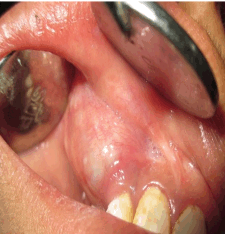
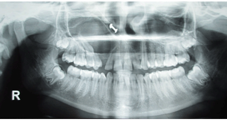
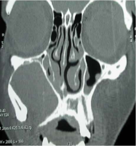
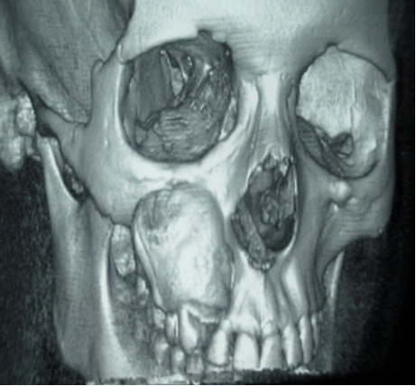
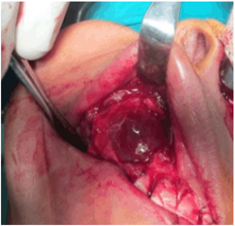
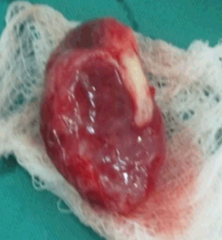
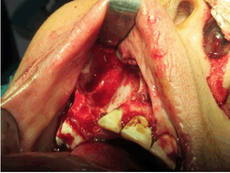
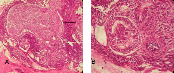
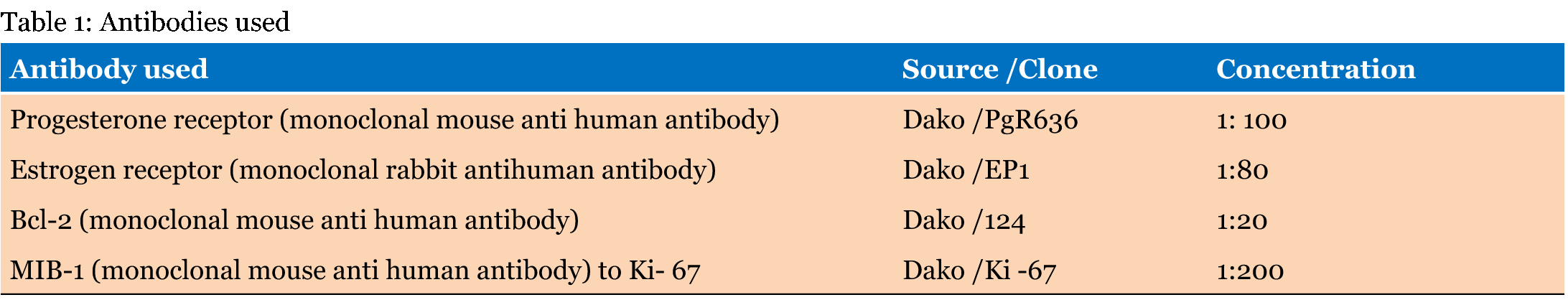
A 20-year-old female presented with a chief complaint of progressive swelling in upper right front region of face since four months. Patient is symptomatic and gave the history of appearance of swelling in last trimester of pregnancy (9th month) since then the swelling has gradually increased in size to the present condition. Patient gave no history of trauma although there was history of pus discharge occasionally from the gingival margin in upper front teeth. On clinical examination, a solitary unilateral swelling was present on upper right front side of the face extending from vermillion border of right upper lip to infra orbital rim measuring about 3x3 cm and causing flaring of the right nostril. Swelling was round in shape with soft to firm in consistency and mild tenderness was elucidated on palpation without any changes in the overlying skin (Figure 1) (Figure 2). On intra oral examination, labial vestibule was obliterated in right upper front teeth due to the swelling, with missing permanent canine and grade 1 mobility of first and second premolars which are tender on percussion without any discoloration. There were no signs of sinus opening or paraesthesia in the same region. As a chair side investigation, fine needle aspiration biopsy was performed which revealed straw colored fluid. A provisional diagnosis of dentigerous cyst associated with impacted canine was made. Carrying the investigations further, OPG and CT scan were advised in order to know the exact extent of the lesion. Orthopantomography revealed a well-defined radiolucency with well corticated borders extending from lateral aspect of root of right maxillary lateral incisor to the mesial aspect of root of 16. The radiolucency encircling the impacted canine and resorption of roots of 1st and 2nd premolars were also noted (Figure 3). CT scan of the face revealed a cyst like lesion associated with impacted canine on right side of the face with thinning of maxillary bone (Figure 4) and (Figure 5). After a through pre-anesthetic evaluation and all the hematological investigations being under normal limits, the lesion was completely enucleated along with impacted canine and extraction of first and second premolars was done under general anesthesia (Figure 6) (Figure 7) (Figure 8). After thorough curettage, the wound was closed using 3-0 vicryl and the specimen was sent for histopathological examination. Macroscopically the lining was attached to cementoenamel junction of impacted canine. Microscopically, section revealed sheets of polyhedral, spindle shaped epithelial cells in nodular arrangement with interspersed eosinophilic material, variable sized duct like structures lined by columnar cells, whorled masses, and rosettes with eosinophilic hyaline masses. There are dispersed calcifications and periphery shows interconnecting strands and cords of dark basophilic cells. The stromal connective tissue surrounding the epithelial component shows delicate to dense collagen. Blood vessels, and reactive bone formation was as seen in Figure 9. Based on histopathological features, a diagnosis of Adenomatoid odontogenic tumor was made. Immunohistochemistry | ||||||
| ||||||
| ||||||
| ||||||
| ||||||
| ||||||
| ||||||
| ||||||
| ||||||
|
| ||||||
| ||||||
|
Discussion
| ||||||
|
Adenomatoid odontogenic tumor is a benign lesion originating from odontogenic epithelium with solid masses of cells and duct like structures in its histological appearance which gives the lesion its name in the true sense. Philipsen and Reichart considered AOT as a non-invasive slow-growing benign lesion [5] while Takahashi et al. through their transferrin detection by immunohistochemistry revealed the neoplastic nature of the tumor [6]. About two-thirds of the cases are diagnosed when the patients are in their twenties, and more than 90%, until they are in their thirties. The ratio of male to female patients with AOT is 1:1.9 [7]. It occurs more frequently in the maxilla than in the mandible with the ratio of frequency being 2.6:1 [8]. The most common site of occurrence is maxillary anterior teeth, usually associated with impacted canines [9]. Clinically, AOT represents slow growing symptom free lesion which may cause the patient to tolerate the swelling for years until it produces an obvious deformity. However, it may cause expansion of bone and displacement of adjacent teeth in advanced stages. It has been hypothetized that the tumor expands in centrifugal pattern initially involving cancellous bone in a linear pattern followed by cortical expansion or resorption [10]. Based on the clinical and topographic features, Philipsen and Reichert have classified AOT into three variants as follicular, extrafollicular and peripheral. The follicular and extrafollicular are intra osseous type of which follicular constitutes 71% of all variants of AOT [7]. The imaging findings of AOT reveal unilocular and well-demarcated cyst-like radiolucency frequently accompanied by impacted teeth. Sometimes multiple radio opacities may be found adjacent to the impacted teeth which are considered to be a characteristic feature of this lesion [11]. A unique feature of AOT that can be observed on CT scan is the presence of radiolucent band surrounding the periphery of the lesion known as capsular space [12]. Under histological examination, the tumor may be partly cystic and in some cases the solid lesion may be present only as masses in the wall of a large cyst. Moreover, eosinophilic non-calcified amorphous material can be found and is called "tumor droplets" which represent electron dense plaques or homogenous matrix [5]. Surgical removal of tumor along with impacted tooth is the mode of treatment. Enucleation of the tumor is reasonably easy as it is well encapsulated. Recurrence rate is also very less as compared to other odontogenic tumors [13]. A variety of intraoral lesions have been associated with pregnancy which includes pyogenic granuloma, focal fibrous hyperplasia or fibroma, peripheral ossifying fibroma and giant cell granulomas [14]. The rapid enlargement of the tumor during pregnancy is attributed to high proliferative activity of tumor cells, inhibition of apoptotic cell loss, or biological characteristics resembling benign odon-togenic tumors under the effect of sex hormones but the underlying mechanism has not been explained in detail by Daley et al. [15]. Estrogen is the sex hormone whose expression significantly increases during pregnancy and is widely studied in this context. It is a sex hormone secreted by the granulosa cells of the ovarian follicle and is essential for the development of internal and external genitalia during the growth period as well as for the reinforcement of the reproductive function during sexual maturity. Estrogen promotes cell growth by binding to nuclear receptors within a cell and translocating into the nucleus. It has been reported that Bcl-2 protein expression is seen in the epithelial tumor cells of all benign odontogenic tumors. It plays an important role in tumor cell survival and regulation of apoptosis by preventing the activation of ICE protease cascade. The tumor cell survival and inhibition of apoptotic cell loss, facilitated by Bcl-2 upon continuous stimulation by estrogen resulted in the enlargement of the tumor during pregnancy [16]. In the present case, the expression level of Bcl-2 is 3+ which indicates strong positive staining. MIB-1L1 is an index of proliferative activity of neoplastic cells determined after counting the number of immunopositive cells per histopathologic field. In the present case, focal localization of Ki-67 has been observed in the neoplastic cells which indirectly determine growth activity of the tumor. Tsukinoki et al. reported that MIB1L1 of Adenomatoid odontogenic tumor in combined epithelial odontogenic tumor was 1.2% [17]. Based on immunohistochemical analysis of the tumor of our case, the tumor cells had weak positive reaction with estrogen but there was strong positive reaction with Bcl-2 which suggests that the rapid tumor growth during pregnancy is facilitated by Bcl-2 upon continuous stimulation by estrogen. | ||||||
|
Conclusion
| ||||||
|
Due to its similar radiological appearance, Adenomatoid odontogenic tumor is often misdiagnosed as a dentigerous cyst. It is often seen in anterior maxillary region, involving an impacted canine. The rate of proliferation of odontogenic tumors during pregnancy is rapid, but definitive treatment can be delayed till parturition or may require immediate attention. Delayed treatment may cause increased morbidity. Enucleation is relatively easy as the tumor is well encapsulated and recurrence is rare. As above mentioned immunohistochemical markers stained positive, a larger case series is needed to definitely implicate growth of odontogenic tumors during pregnancy. | ||||||
|
References
| ||||||
| ||||||
|
[HTML Abstract]
[PDF Full Text]
|
|
Author Contributions
G. Siva Prasad Reddy – Substantial contributions to conception and design, Acquisition of data, Analysis and interpretation of data, Drafting the article, Revising it critically for important intellectual content, Final approval of the version to be published G.V. Reddy – Analysis and interpretation of data, Revising it critically for important intellectual content, Final approval of the version to be published N.V.S. Sekhar Reddy – Analysis and interpretation of data, Revising it critically for important intellectual content, Final approval of the version to be published K. Sravan Kumar Reddy – Analysis and interpretation of data, Revising it critically for important intellectual content, Final approval of the version to be published Rehana Sulthana – Analysis and interpretation of data, Revising it critically for important intellectual content, Final approval of the version to be published Phanitej G. – Analysis and interpretation of data, Revising it critically for important intellectual content, Final approval of the version to be published E. Divya Jyothi – Analysis and interpretation of data, Revising it critically for important intellectual content, Final approval of the version to be published |
|
Guarantor of submission
The corresponding author is the guarantor of submission. |
|
Source of support
None |
|
Conflict of interest
Authors declare no conflict of interest. |
|
Copyright
© 2016 G. Siva Prasad Reddy et al. This article is distributed under the terms of Creative Commons Attribution License which permits unrestricted use, distribution and reproduction in any medium provided the original author(s) and original publisher are properly credited. Please see the copyright policy on the journal website for more information. |
|
|