| |
|
|
|
Clinical Image
| ||||||
| Pneumopericardium resulting in pneumoperitoneum in a newborn with congenital diaphragmatic hernia | ||||||
| Thomas Pennaforte1, Antoine Payot2 | ||||||
|
1MD, Department of Pediatrics, Neonatology, Sainte-Justine University Health Center, Université de Montreal, Montreal, Quebec, Canada.
2MD, PhD, Department of Pediatrics, Neonatology, Sainte-Justine University Health Center, Université de Montreal, Montreal, Quebec, Canada. | ||||||
| ||||||
|
[HTML Abstract]
[PDF Full Text]
[Print This Article]
[Similar article in Pumed] [Similar article in Google Scholar] 
|
| How to cite this article |
| Pennaforte T, Payot A. Pneumopericardium resulting in pneumoperitoneum in a newborn with congenital diaphragmatic hernia. Int J Case Rep Images 2016;7(9):615–617. |
|
Case Report
| ||||||
|
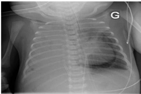
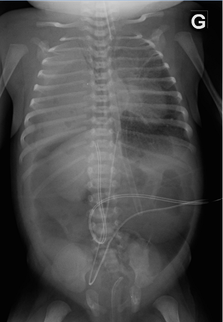
A five-day old girl presented with a pneumopericardium discovered fortuitously on a radiograph (Figure 1). She was born at term with a prenatal diagnosis of right-sided congenital diaphragmatic hernia (CDH) and required high frequency ventilation and inotrope/pressor therapy since birth. On day-6 the pneumopericardium increased and a pneumomediastinum was noted (Figure 2). The newborn remained stable without signs of cardiac tamponade. On day-7, the pneumopericardium/mediastinum resolved and a pneumoperitoneum appeared (Figure 3). The condition of the child improved at the same time and it was possible to gradually diminish ventilation pressures and discontinue inotrope/pressor therapy. No intestinal perforation was noted during surgery a few hours later. | ||||||
| ||||||
| ||||||
|
| ||||||
| ||||||
|
Discussion
| ||||||
|
Neonatal pneumopericardium is a rare condition in the neonate, usually caused by mechanical ventilation [1]. The only previously reported association of pneumopericardium with pneumomediastinum and pneumoperitoneum was in a 77-year-old patient who received artificial ventilation following blunt chest trauma [2]. We believe that the air in the pericardial space dissected into the thoracoabdominal peritoneal cavity leading to hemodynamic and respiratory improvement in this newborn with CDH. | ||||||
|
Conclusion
| ||||||
|
This is the first case of a newborn with CDH presenting with pneumopericardium resulting in pneumoperitoneum and improvement of respiratory and hemodynamic functions before surgery. Keywords: Congenital diaphragmatic hernia (CDH), Pneumopericardium, Pneumomediastinum | ||||||
|
References
| ||||||
| ||||||
|
[HTML Abstract]
[PDF Full Text]
|
|
Author Contributions
Thomas Pennaforte – Substantial contributions to conception and design, Acquisition of data, Analysis and interpretation of data, Drafting the article, Revising it critically for important intellectual content, Final approval of the version to be published Antoine Payot – Analysis and interpretation of data, Revising it critically for important intellectual content, Final approval of the version to be published |
|
Guarantor of submission
The corresponding author is the guarantor of submission. |
|
Source of support
None |
|
Conflict of interest
Authors declare no conflict of interest. |
|
Copyright
© 2016 Thomas Pennaforte et al. This article is distributed under the terms of Creative Commons Attribution License which permits unrestricted use, distribution and reproduction in any medium provided the original author(s) and original publisher are properly credited. Please see the copyright policy on the journal website for more information. |
|
|