  |

|
 |
|
Case Report
| ||||||
| A fatal case of empyema due to Streptococcus intermedius associated pneumonia masquerading as acute pancreatitis in an otherwise healthy middle-aged woman | ||||||
| Anthea B. Mahesan Paul1, Lary Simms1, Abraham Ebenezer Paul1, Jojo Yorke1, Christopher Schmidseder1 | ||||||
|
1Clark County Coroners Office, 1704 Pinto Lane, Las Vegas, Nevada, USA.
| ||||||
| ||||||
|
[HTML Abstract]
[PDF Full Text]
[Print This Article]
[Similar article in Pumed] [Similar article in Google Scholar] 
|


| How to cite this article |
| Paul ABM, Simms L, Paul AE, Yorke J, Schmidseder C. A fatal case of empyema due to Streptococcus intermedius associated pneumonia masquerading as acute pancreatitis in an otherwise healthy middle-aged woman. Int J Case Rep Images 2016;7(8):524–528. |
|
Abstract
|
|
Introduction:
Members of the S. milleri group Streptococcus intermedius, Streptococcus anginosus, and Streptococcus constellatus intermedius, are important pathogens implicated in respiratory illnesses such as bacterial pneumonia, pulmonary abscesses and empyema. Diagnosis of respiratory illnesses is largely dependent on accurate identification of symptomology and physical exam as radiological evidence is not always present.
Case Report: In this case report, we present a fatal case of Streptococcus intermedius associated pneumonia causing empyema with an unusual constellation of symptoms and laboratory tests masquerading as acute pancreatitis in an otherwise healthy middle-aged woman. Conclusion: Our case adds to literature a novel clinical and laboratory presentation of S. intermedius empyema and the potentially fatal ramifications if not accurately diagnosed in an otherwise healthy woman. | |
|
Keywords:
Empyema, Pancreatitis, Pneumonia, Streptococcus intermedius
| |
|
Introduction
| ||||||
|
Streptococcus milleri was first named in 1956 by O. Guthof when describing non-haemolytic species of commensal streptococci found in the oral cavity [1]. It has since been found as a normal commensal bacterium of the mouth, appendix, vaginal secretions and among fecal flora in 16–67% of healthy adults [2] [3]. The Streptococcus milleri group are anerobic gram-positive cocci in pairs or chains and contains three members, Streptococcus intermedius, Streptococcus anginosus, and Streptococcus constellatus intermedius. Members of the S. milleri group are important pathogens implicated in respiratory illnesses such as bacterial pneumonia, pulmonary abscesses and empyema [5] [9]. Diagnosis of respiratory illnesses is largely dependent on accurate identification of symptomology and physical exam as radiological evidence is not always present. Of the symptomology associated with pneumonia, the most common respiratory symptoms are cough, dyspnea, and sputum production, whereas the most frequently reported non-respiratory symptoms are fatigue, fever, and chills [6]. In this case report, we present a fatal case of Streptococcus intermedius associated pneumonia causing empyema with an unusual constellation of clinical and laboratory symptoms masquerading as acute pancreatitis in an otherwise healthy middle-aged woman. | ||||||
|
Case Report
| ||||||
|
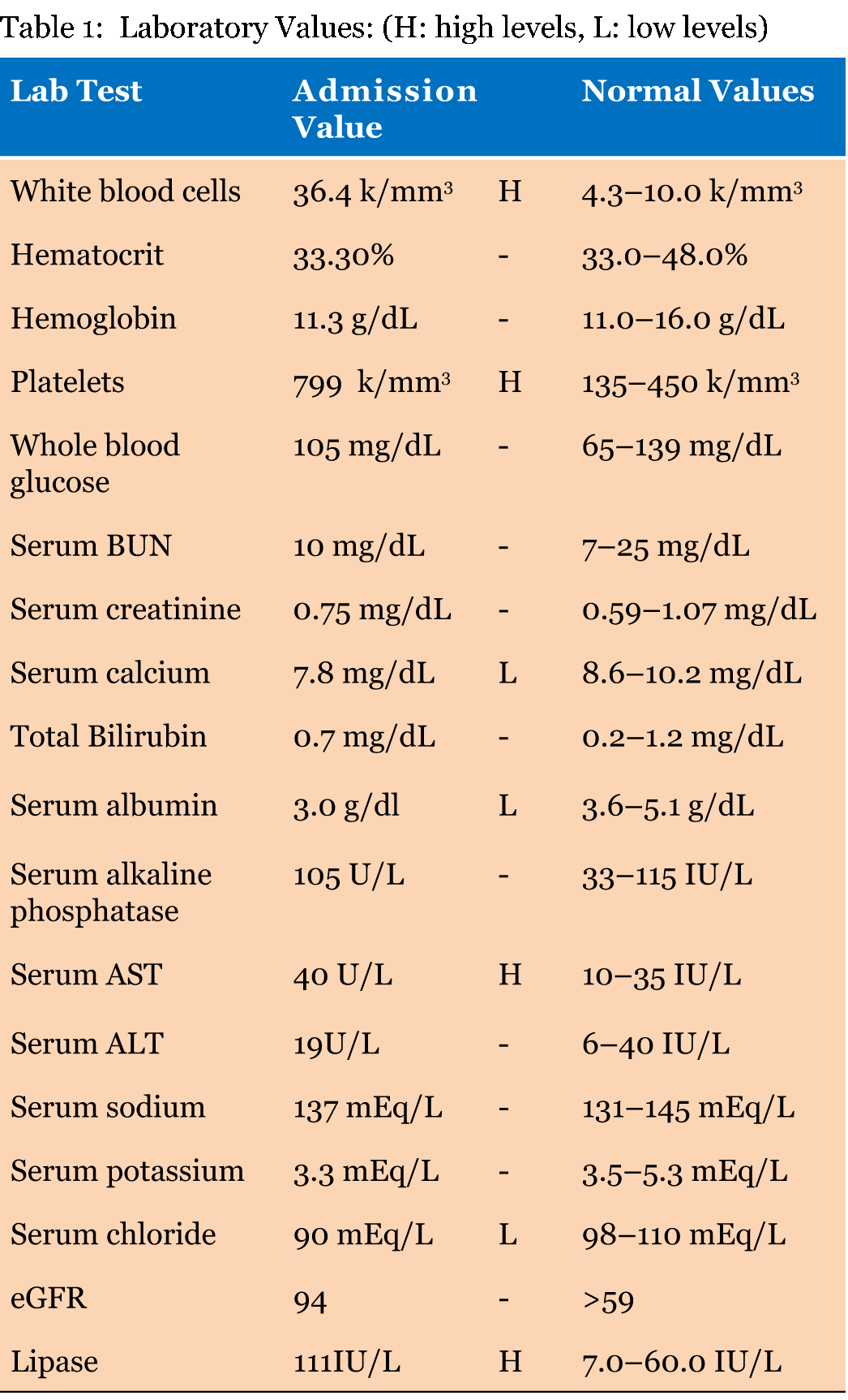
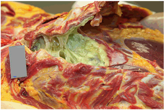
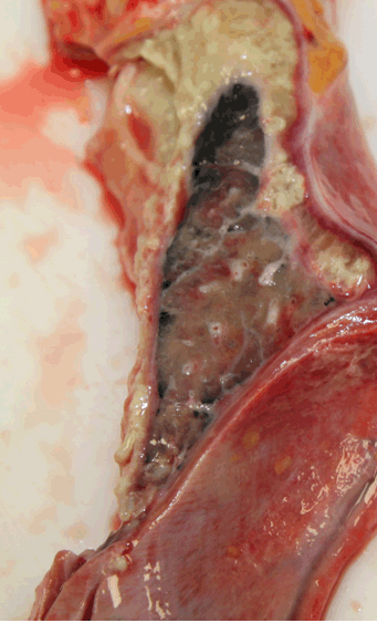
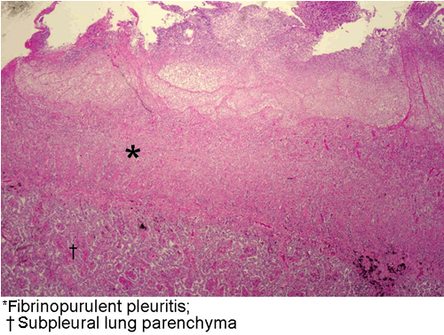
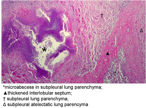
A 48-year-old Caucasian female presented to the emergency room with severe sharp non-radiating epigastric pain, associated with, coryza, nausea, diarrhea with generalized myalgia. The nonspecific symptoms began 10 days prior and gradually increased in severity despite over counter cough-and-cold medication. The patient was in good overall health with a medical history of anxiety, chronic lumbar back pain and lumbar laminectomy four years ago. She was taking both pain and anti-anxiety medication. The patient was a non-smoker and admitted occasional alcohol use. Vital signs on admission are temperature 98.5°C, blood pressure 106/59 mmHg, heart rate 114 bpm and respiratory rate 18/min. Pulse oximetry showed 96% oxygen saturation. Physical examination revealed tachycardia (114 bpm), a soft, tender abdomen and coarse crackles in the right lung field. Laboratory findings on admission showed significant leukocytosis (36.4 k/mm3), thrombocytosis (799 k/mm3), and mildly elevated lipase levels (111 IU/L) (Table 1). Our patient was admitted with the initial diagnosis of suspected acute pancreatitis and was started on IV fluids. Unfortunately, against medical advice the patient discharged herself before other confirmatory tests and treatment could have been given. She was given anti-inflammatories for pain and asked to return to the emergency room as soon as possible for re-admission. At home she continued to experience nonspecific symptoms until she was found deceased in bed three days later. At autopsy, the external examination was unremarkable. Upon removing the breastplate, the right pleural cavity exhibited a large empyema composed of loculated purulent exudate around 300 ml in estimated volume (Figure 1). Removal and dissection of the right lung demonstrated the loculated exudate was firmly adherent to the visceral pleural surface (Figure 2). Postmortem microbiology findings showed negative urine bacterial antigens of Group B Strep, H. influenzae, Streptococcus pneumoniae and N. meningitidis. Two different right pleural cultures were positive for Streptococcus intermedius; all other postmortem viral and bacterial tests were non-contributory. Microscopic examination of autopsy tissues showed acute fibrinopurulent inflammation of the surface of the right lung (Figure 3) associated with areas of necrosis and repair. The underlying lung showed broad areas of alveolitis and repair with scattered microabscesses, (Figure 4) and some small airways exhibited necrosis. Microscopic examination of the pancreas had normal number and appearance of islets of Langerhans with normal surrounding acinar tissue. The rest of the autopsy was unremarkable. The final autopsy diagnosis was empyema of the right chest due to Streptococcus intermedius complicated by necrotizing pneumonia. | ||||||
| ||||||
| ||||||
| ||||||
| ||||||
| ||||||
|
Discussion
| ||||||
|
Though the Streptococci milleri group is morphologically similar they appear to be associated with different clinical diseases and regions of the body. Whiley et al. found Streptococcus anginosus was the most prevalent of the three S. milleri bacteria accounting for 59% (89/151) of infections, most commonly found in the gastrointestinal tract and genitourinary tract [7]. Streptococcus constellatus intermedius accounted for 23% (33/151) of all S. milleri infections and was primarily found in the respiratory tract [8]. Streptococcus intermedius was only found in 19% (29/151) of Streptococcus milleri infections and was present most commonly in 86% (18/21) of head and neck infections [8]. In the respiratory tract, S. intermedius was found to be responsible for only 14% (1/12) of cases causing infection [8]. In literature, infections caused by S. milleri compared to those by Streptococcus pneumonia seem to affect hosts with underlying medical conditions and present with a longer course [9]. Of the five fatalities reported by Wong et al. with S. milleri associated empyema, all five had significant comorbid disease such as malignancy, invasive procedures, stroke, or dental disease [9]. This, however, was not the case in our patient who was in generally good health with no significant medical conditions, or recent procedures. Our case adds to literature the potential fatal ramification of infection by S. milleri if not accurately diagnosed in an otherwise healthy woman. Respiratory symptomology associated with pneumonia has been reported to include cough, dyspnea, sputum production, pleuritic chest pain, and hemoptysis [7][9]. Non-respiratory symptoms that have been reported with pneumonia have ranged from fatigue, fever, chills, anorexia, sweats, coryza, headache, earache, myalgia, nausea, sore throats, vomiting, diarrhea, and abdominal pain [7] [9]. Our patient exhibited many of the documented symptoms in literature, however, interestingly in addition had elevated lipase levels (111 IU/L) which may have influenced the clinician to suspect acute pancreatitis. Elevated lipase levels have been reported in pancreatic etiology such as pancreatitis and in obstruction of the pancreatic duct, as well as in non-pancreatic etiology such as renal disease, alcoholism, diabetic ketoacidosis, post-endoscopic retrograde cholangiopancreatography and gastrointestinal diseases such as acute cholecystitis, nonspecific diarrhea, bowel obstruction, intestinal infarction, duodenal ulcers, and liver diseases [10]. A study by Tetrault found that isolated lipase levels were increased in 38/62 (61%) of patients with gastrointestinal disorders, specifically they found that 9/38 (23/6%) of patients with gastrointestinal disease and an isolated elevated lipase level had nonspecific diarrhea, as was the case in our patient [10]. In retrospect, we attribute the elevated lipase levels in our patient to her nonspecific diarrhea, as autopsy histopathology of all other organs, including the pancreas were unremarkable. Empyema with clinical diagnosis has a very high mortality rate ranging between between 6–24% [4]. Though the symptoms exhibited by our patient are listed in the reported symptomology in literature, this constellation of symptoms was unusual in the circumstances presented. Our patient did exhibit significant relevant respiratory findings to warrant further investigation, however, the constellation of abdominal pain, elevated lipase levels, and diarrhea misled the diagnostic team to the incorrect initial diagnosis. We feel with patient cooperation, appropriate hospitalization and necessary diagnostic imaging and laboratory tests; our patient would have been appropriately diagnosed and treated. Our case demonstrates the need for awareness of the vast range of symptoms related to empyema by Streptococcus intermedius associated pneumonia to prevent further morbidity and mortality caused by this disease. | ||||||
|
Conclusion
| ||||||
|
Our case adds to literature a novel clinical and laboratory presentation of S. intermedius empyema and the potentially fatal ramifications if not accurately diagnosed in an otherwise healthy woman. | ||||||
|
Acknowledgements
| ||||||
|
We would like to thank the Clark County Coroner's Office for the ongoing support. | ||||||
|
References
| ||||||
| ||||||

|
[HTML Abstract]
[PDF Full Text]
|
|
Author Contributions
Anthea B. Mahesan Paul – Substantial contributions to conception and design, Acquisition of data, Analysis and interpretation of data, Drafting the article, Revising it critically for important intellectual content, Final approval of the version to be published Lary Simms – Analysis and interpretation of data, Revising it critically for important intellectual content, Final approval of the version to be published Abraham Ebenezer Paul – Analysis and interpretation of data, Revising it critically for important intellectual content, Final approval of the version to be published Jojo Yorke – Analysis and interpretation of data, Revising it critically for important intellectual content, Final approval of the version to be published Christopher Schmidseder – Analysis and interpretation of data, Revising it critically for important intellectual content, Final approval of the version to be published |
|
Guarantor of submission
The corresponding author is the guarantor of submission. |
|
Source of support
None |
|
Conflict of interest
Authors declare no conflict of interest. |
|
Copyright
© 2016 Anthea B. Mahesan Paul et al. This article is distributed under the terms of Creative Commons Attribution License which permits unrestricted use, distribution and reproduction in any medium provided the original author(s) and original publisher are properly credited. Please see the copyright policy on the journal website for more information. |
|
|