  |

|
 |
|
Case Report
| ||||||
| Onlay mesh repair for spontaneous lumbar hernia: A case report | ||||||
| Biswal Jayanta Kumar1, Mahapatra Tanmaya2, Guria Sourabh2, Supreet Kumar2, Meher Dibyasingh2, Kanhat Karesh Samu2 | ||||||
|
1MS, Associate Professor, Department of General Surgery, S.C.B. Medical College, Cuttack, Odisha, India.
2Postgraduate, Department of General Surgery, S.C.B. Medical College, Cuttack, Odisha, India. | ||||||
| ||||||
|
[HTML Abstract]
[PDF Full Text]
[Print This Article]
[Similar article in Pumed] [Similar article in Google Scholar] 
|


| How to cite this article |
| Biswal JK, Tanmaya M, Sourabh G, Supreet K, Dibyasingh M, Kanhat KS. Onlay mesh repair for spontaneous lumbar hernia: A case report. Int J Case Rep Images 2016;7(7):481–485. |
|
Abstract
|
|
Introduction:
Lumbar hernia is a rare hernia. It herniates through the superior lumbar triangle (Grynfeltt-Lesshaft triangle) or inferior lumbar triangle (Petit triangle). It can be classified as congenital or acquired, which may be primary or secondary.
Case Report: A 70-year-old male with a reducible left sided superior lumbar hernia. Intraoperatively, there was a small defect and it was repaired with primary closure and an onlay meshplasty. The patient was absolutely asymptomatic during the follow-up. Conclusion: Though there are many techniques for repair of lumbar hernia, in case of a small defect, a primary repair with a tension free onlay meshplasty can be a quick procedure with good result. | |
|
Keywords:
Flank swelling, Grynfeltt-Lesshaft triangle, Inferior lumbar triangle, Lumbar hernia, Onlay mesh repair, Superior lumbar triangle
| |
|
Introduction
| ||||||
|
Lumbar hernia is one of the rarest forms of abdominal wall hernias with only about 300 cases of primary lumbar hernias being reported over last four centuries [1]. First described by Barbette in 1672, the existence of this variant of hernia is known for four centuries and the first case was reported by Garangoet in 1731 [2]. | ||||||
|
Case Report
| ||||||
|
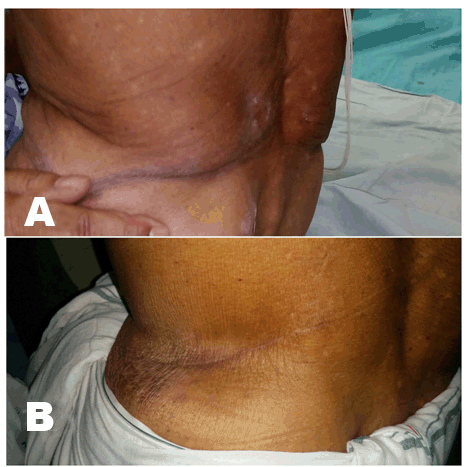
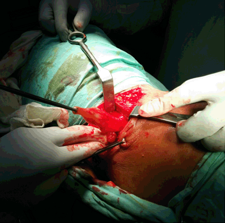
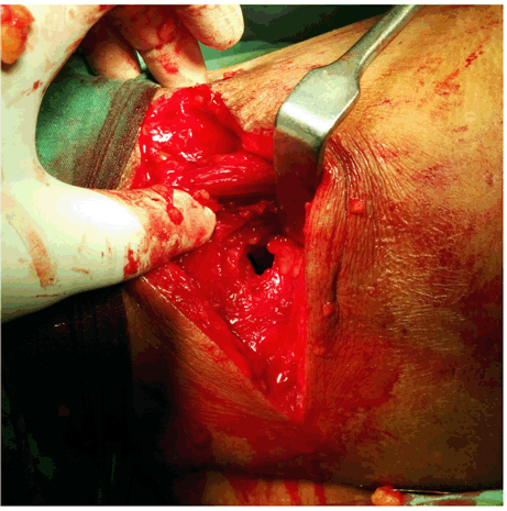
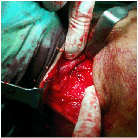
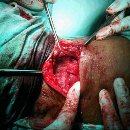
A 70-year-old male patient presented with gradually increasing left flank swelling of duration one and half years (Figure 1A). Over the past six months there was local discomfort and dull pain. There was no history of trauma, previous surgery, hematuria or altered bowel habits. On examination there was a globular, soft, non-tender, reducible, non-pulsatile swelling of size 7×5 cm located in the left superior lumbar triangle with an expansile cough impulse. The swelling was more prominent with straining or a standard Valsalva maneuver and disappearing with prone position The examination of rest of the abdomen, right flank, back & hernia orifices were normal. Ultrasonography revealed dehiscence of abdominal wall of size 15 mm in the left lumbar region. So a diagnosis of left sided reducible lumbar hernia was made on the basis of clinical and radiological finding and the patient was taken up for surgery. The patient was placed in modified right lateral decubitus position. Oblique left lumbar incision was given. The hernia sac was identified and opened. The content being the extra peritoneal fat was partially excised and the rest was reduced (Figure 2). The size of the defect was about 1.5 cm (Figure 3) which was primarily repaired with (1–0) polypropylene interrupted suture (Figure 4) and then an onlay polypropylene mesh was given over the defect (Figure 5). The skin was closed with a negative suction drain. The postoperative periods were uneventful and the patient was discharged on 4th postoperative day on analgesics. During the follow-up visit after five months the operation scar was found to have healed well and the patient was absolutely asymptomatic (Figure 1B). | ||||||
| ||||||
| ||||||
| ||||||
| ||||||
| ||||||
|
Discussion
| ||||||
|
The lumbar region is bordered by the twelfth rib superiorly, the iliac crest inferiorly, the erector spinae muscles of the back posteriorly, and a vertical line between the anterior tip of the twelfth rib and the iliac crest anteriorly. The region contains two anatomic triangles, through which the rare lumbar hernia can form. The inferior lumbar triangle of Petit is the more common of the two. Its anterior border is the posterior edge of the external oblique muscle, the posterior border is the anterior extent of the latissimus dorsi muscle, and the inferior border is the iliac crest. Lumbar triangle of Grynfeltt, also known as the superior lumbar triangle is bounded by the twelfth rib and the serratus posterior inferior muscle, the posterior border of the internal oblique muscle, and by the quadratus lumborum and erector spinae muscles posteriorly. The floor of the superior triangle is composed of transversalis fascia and the entire triangular space is covered posteriorly by the latissimus dorsi muscle [3]. Lumbar hernias have been classified as congenital (20%) or acquired (80%). An acquired hernia may be primary or secondary. Secondary lumbar hernias are of traumatic or post-surgical (flank incisions, renal surgery, iliac bone harvesting) etiology comprising about 25% of acquired hernias [4]. Another way of classifying lumbar hernia is on the basis of content, they are of two types: extraperitoneal hernia with no sac, containing only fat or sliding retroperitoneal organs (paraperitoneal), and peritoneal hernia that may include intraperitoneal organs such as small bowel, omentum, ovary and stomach [5]. In most of the times the patients are usually asymptomatic, but sometimes may complain of backache, flank pain or a dragging sensation. These hernias are known to have painless progressive enlargement in size [6]. The differential diagnosis includes lipoma, soft tissue tumors, hematoma or an abscess. In obese patients detection of a mass is usually difficult. It is observed that incidences of bowel incarceration may occur in 25% but strangulation is rare because of wide hernial neck [7]. Ultrasonography or computed tomography (CT) imaging are usually obtained in patients with suspected lumbar hernia to confirm the diagnosis [3]. Computed tomography scan is the diagnostic modality of choice. Computed tomography scan is able to delineate muscular and fascial layers, a defect in one or more of these layers, and the presence of herniated fat and/or viscera [4]. Surgical repair is the treatment of choice. Repair can be done either open or endoscopically. In open technique, after reduction of the contents, if the sac is found to be narrow, exploration with ligation of the sac is warranted. A sac with wide neck can be inverted and plicated. The defect is managed according to its size and the status of tissues around it: one of the strategies is to suture it primarily with interrupted heavy non-absorbable sutures. For larger defects some authors postulate the use of tensor fascia lata rotational flaps or free fascial grafts. Nowadays in the era of meshplasty a non-absorbable mesh is usually preferred for reconstruction. Depending on the size of the defect, it can be placed as an onlay, inlay or underlay [8]. The onlay technique involves primary closure of the fascia defect and placement of a mesh over the anterior fascia. Inlay repair involves securing the mesh to the fascial edge without overlap, whereas underlay repair involves placing the mesh below the fascial components. In our case, as the defect was small, it was primarily repaired with non-absorbable interrupted suture and to strengthen it an onlay tension free meshplasty was done with a polypropylene mesh. Recently, minimally invasive approaches to repair of lumbar hernias have been reported. These involve either intraperitoneal laparoscopy necessitating takedown of the lateral peritoneal reflection of the colon to facilitate exposure of the hernia defect [9], or retroperitoneoscopy in which the lateral retroperitoneal space is entered and insufflated [10]. | ||||||
|
Conclusion
| ||||||
|
Primary lumbar hernia is a rare clinical entity and needs a high index of suspicion during day to day practice. A good history, general physical and radiological examination can rule out most of the differential diagnoses. Strengthening the defect can be done with synthetic mesh either open or endoscopic approach. With a small defect, open approach with primary closure and onlay tension free mesh repair can be a quick procedure with good result. | ||||||
|
References
| ||||||
| ||||||

|
[HTML Abstract]
[PDF Full Text]
|
|
Author Contributions
Jayanta Kumar Biswal – Substantial contributions to conception and design, Acquisition of data, Analysis and interpretation of data, Drafting the article, Revising it critically for important intellectual content, Final approval of the version to be published Tanmaya Mahapatra – Substantial contributions to conception and design, Acquisition of data, Analysis and interpretation of data, Revising it critically for important intellectual content, Final approval of the version to be published Sourabh Guria – Acquisition of data, Analysis and interpretation of data, Revising it critically for important intellectual content, Final approval of the version to be published Supreet Kumar – Acquisition of data, Analysis and interpretation of data, Revising it critically for important intellectual content, Final approval of the version to be published Dibyasingh Meher – Acquisition of data, Drafting the article, Final approval of the version to be published Kanhat Karesh Samu – Acquisition of data, Drafting the article, Final approval of the version to be published |
|
Guarantor of submission
The corresponding author is the guarantor of submission. |
|
Source of support
None |
|
Conflict of interest
Authors declare no conflict of interest. |
|
Copyright
© 2016 Jayanta Kumar Biswal et al. This article is distributed under the terms of Creative Commons Attribution License which permits unrestricted use, distribution and reproduction in any medium provided the original author(s) and original publisher are properly credited. Please see the copyright policy on the journal website for more information. |
|
|
|
About The Authors
| |||
| |||
| |||
| |||
| |||
| |||
| |||


