|

|
 |
|
Case Report
| ||||||
| A giant omental cyst mimicking ascites in a three-year-old Congolese child | ||||||
| Léon Tshilolo1,2, Rudy Lukamba1, Alphonse Simbi1, Valentin Kazadi3, Christian Mayemba1,4, Bienvenu Lebwaze4, Raphael Kalengayi4 | ||||||
|
1MD, Paediatrics, Centre Hospitalier Monkole.
2MD, Centre de Formation et d'Appui Sanitaire (CEFA). 3MD, Surgery Department, Cliniques Universitaires de Kinshasa. 4MD, Department of medical Biology, University of Kinshasa). | ||||||
| ||||||
|
[HTML Abstract]
[PDF Full Text]
[Print This Article]
[Similar article in Pumed] [Similar article in Google Scholar] 
|


| How to cite this article |
| Tshilolo L, Lukamba R, Simbi A, Kazadi V, Mayemba C, Lebwaze B, Kalengayi R. A giant omental cyst mimicking ascites in a three-year-old Congolese child. Int J Case Rep Images 2016;7(7):471–475. |
|
Abstract
|
|
Introduction:
Omental and mesenteric cysts are rare intra-abdominal lesions. They can present as an abdominal distension and make differential diagnosis with ascites.
Case Report: A three-year-old boy with fever, under nutrition and abdominal distension previously diagnosed as ascites was submitted to laparotomy. We founded a large multi lobed cyst from the large omentum filling almost the entire abdominal cavity and containing sero-hemorrhagic to very dark liquid. The histopathologic examination showed a cystic formation lined by cubic mesothelium cells, with abundant inflammatory infiltrate rich in lymphocytes and histiocytes, mixed with a few giant cells of foreign body of melanotic origin. Conclusion: The diagnostic role of sonography in abdominal distension of unknown etiology is emphasized. | |
|
Keywords:
Ascites, Child, Melanotic pigments, Omental cyst
| |
|
Introduction
| ||||||
|
Omental and mesenteric cysts are rare intra-abdominal lesions which may be congenital, traumatic, neoplastic or infectious in origin [1] [2]. The similarity of his clinic with abdominal masses, intra-peritoneal as extra peritoneal, as well as intra-abdominal effusions (ascites), makes the diagnosis somewhat difficult [3]. The preoperative diagnosis can be made by the combined modalities of ultrasound and CT scan [2] [4]. We report the case of a three-year-old child admitted for abdominal ascites and under nutrition who underwent to laparotomy. The role of multidisciplinary approach and diagnostic tools is emphasised. | ||||||
|
Case Report
| ||||||
|
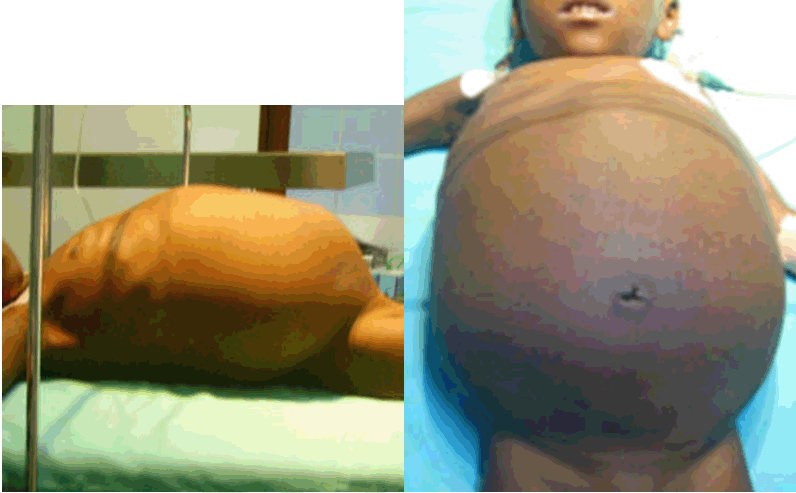
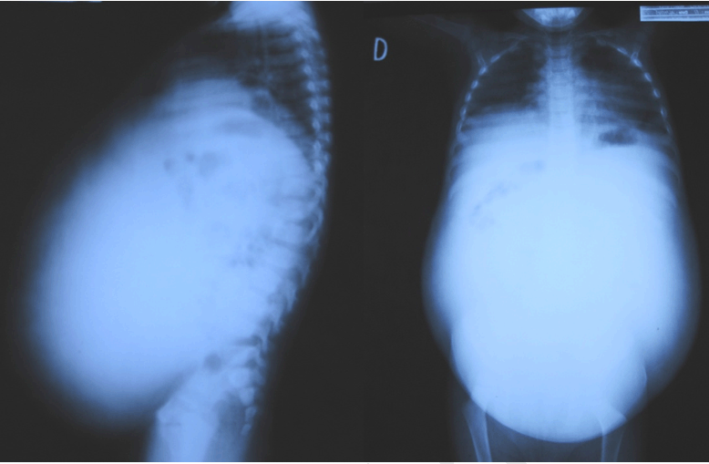
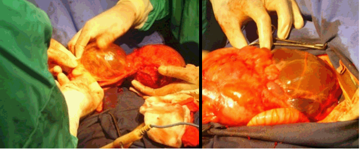
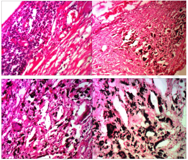
A three-year-old boy was first admitted three months before to another hospital in Kinshasa and then referred to Monkole Hospital Centre for evaluation of abdominal distension and fever lasting for two weeks. The patient was a neglected child who was welcomed in a charity residence in Kinshasa. No available data was known about his previous medical history. In the previous admission, he was submitted to a malaria treatment and a paracentesis was performed revealing an apparent exsudative fluid. No biological data were available. On physical examination, the child was undernourished, pale and febrile (39°C). The heart beats were rapid at 140 beats/min without abnormal murmur; respiratory rate varied from 35–40 cycles/min and the pulmonary sounds were reduced at the bottom. The abdomen was very protuberant and scarcely tender, with a positive fluid thrill and a vaguely "no well-defined" mass in the right hypochondria and pubic region. The other abdominal organs (liver and spleen) were not easy to appreciate. A venous collateral circulation and tattoos were noted on the abdominal wall. The abdomen circumference measured 57 cm (Figure 1). Significant biological parameters at admission were: white blood cell count 13,000/mm3 (N49 L49 M0 E2), hemoglobin 9.8 g/dl, hematocrit 26.9%, MCV 70 fl; platelets 579,000/mm3. ESR 118/h. Quick time: 80% and INR1.2. Rapid HIV test and tuberculosis test were negative. Blood urea, electrolytes and liver and kidney function studies were within normal limits. Chest and abdominal X-ray showed no lesions in the lung parenchyma, but more horizontally ribs and more upper right diaphragm. Abdomen was fulfilled by an opaque homogenous mass with concomitant displacement of the intestines to posterior and upper area. No calcification was observed (Figure 2). Abdominal ultrasound revealed the presence of a large cystic mass with multiple septa and mobile internal echoes (brown movements) from epigastrium to pelvis (Figure 3). All the solid organs (liver, spleen, kidneys and pancreas) were normal but displaced by the mass. There was no ascites. Diagnosis of "mesothelium tumoral mass" or "peritoneal myxoma" was evoked. A selective laparotomy was then indicated. The exploration of the abdominal cavity revealed a solid and multiseptated cystic mass originated from the large omentum, adjacent to the liver (Figure 4). The content of the mass was a liquid variable from sera aspect in one cavity and to very dark aspect in the others. The mass was completely excised and the postoperative course was uneventful. There has been no recurrence till now after five years of the laparotomy. Histological examination showed a fibrous tissue having the shape of the wall of a tortuous cystic. Some fragments were the seat of a nonspecific chronic inflammation with the presence of giant cells of foreign body. There was no presence of cartilage tissue in the fragments or malignancy or any neoplasm. The overall look was pleading for an inflamed and remodeled cystic mass whose nature could be an inflamed cyst enterogene and reworked or other congenital hamartoma cyst kind overhauled. Proofreading blades noted cystic formation lined by cubic mesothelium cells, sometimes flattened, and supported by a fibro-fatty tissue that was the seat of vascular congestion with hemorrhagic suffusion. No significant lymphatic vessels proliferation has been observed. Abundant inflammatory infiltrate rich in lymphocytes and histiocytes was also present, mixed with a few giant cells of foreign body, next to brown-black deposits reminiscent of melanin pigments. The special coloration Fontana confirmed the nature of the melanin pigments (Figure 5). The Perls coloration was negative, excluding the ferric deposits. Diagnosis of inflamed melanotic peritoneal cyst was selected. | ||||||
| ||||||
| ||||||
| ||||||
| ||||||
|
| ||||||
|
Discussion
| ||||||
|
Mesenteric, omental, and retroperitoneal cysts are unusual causes of intra-abdominal masses in childhood. Mesenteric cysts in children are mostly lymphatic malformations of mesentery or omentum origin and clinical presentation varies from abdominal distension, chronic abdominal pain, subocclusion to sudden onset of peritonitis or volvulus [2] [5]. Different developmental theories for these cysts are discussed, including the benign proliferation of the ectopic mesenteric lymphatics that do not communicate with the rest of the lymphatic system, the failure to join the embryonic lymphatic spaces to the venous system, the failure of the fusion of mesenteric leaves, the deficiency of the normal lymphaticovenous shunts, trauma, neoplasm, and the localized degeneration of lymph nodes [2] [5]. Histologically, the nature of the cyst is determined according to the type of epithelium that lines the inner layer of the wall. Lymphangiomas are the most common cause of mesenteric and omental cysts comprising 90% of cases and the remaining cysts are mesothelial cysts. Thus, the peritoneal cysts are divided into several types and their origin is not currently well understood [6]. Melanotic peritoneal cyst multilocular has been described as a particular entity of peritoneal cysts which likely represent a peritoneal inclusion cyst related to chronic inflammation [7]. The pseudomelanosis coli is another possibility that could explain the presence of melanin pigments in the cyst wall. It is a benign condition due to the accumulation of blackish-brown pigment lipofuscinic kind in the lamina propria of the colon, with the possibility of migration [8]. It is often due to the abuse of laxatives such as anthraquinone derivatives. The brown-black deposits correspond to cellular apoptotic bodies phagocytized by intraepithelial macrophages migrate to the lamina propria after being transformed into lipofuscinic pigments by lysosomes [9]. Our patient had a giant cyst compressing the intestines with a possible constipation, but with no history of abuse of laxatives. The presence of hemorrhagic fluid has been described in omental cysts but only few cases reported the histological description. In this case report, the Perls coloration was negative for the ferric deposits and the presence of melanin pigments in the cyst wall has no clear explanation. The presence of lipofuscin pigments would be an event that would complicate a cyst whose genesis is not clearly defined. The clinical history of abdominal distension with absence of any well demarcated mass can easily make the diagnosis of ascites as described in previous case reports [3] [5][10]. | ||||||
|
Conclusion
| ||||||
|
A multidisciplinary approach involving pediatricians, radiologists and surgeons will be encouraged in order to avoid abusive paracentesis in children with abdominal distention mimicking ascites. Clinical diagnosis of omental cyst is not evident and required the ultrasound exploration before the laparoscopic excision. | ||||||
|
References
| ||||||
| ||||||


|
[HTML Abstract]
[PDF Full Text]
|
|
Author Contributions
Léon Tshilolo – Substantial contributions to conception and design, Acquisition of data, Analysis and interpretation of data, Drafting the article, Revising it critically for important intellectual content, Final approval of the version to be published Rudy Lukamba – Analysis and interpretation of data, Revising it critically for important intellectual content, Final approval of the version to be published Alphonse Simbi – Analysis and interpretation of data, Revising it critically for important intellectual content, Final approval of the version to be published Valentin Kazadi – Analysis and interpretation of data, Revising it critically for important intellectual content, Final approval of the version to be published Christian Mayemba – Analysis and interpretation of data, Revising it critically for important intellectual content, Final approval of the version to be published Bienvenu Lebwaze – Analysis and interpretation of data, Revising it critically for important intellectual content, Final approval of the version to be published Raphael Kalengayi – Analysis and interpretation of data, Revising it critically for important intellectual content, Final approval of the version to be published |
|
Guarantor of submission
The corresponding author is the guarantor of submission. |
|
Source of support
None |
|
Conflict of interest
Authors declare no conflict of interest. |
|
Copyright
© 2016 Léon Tshilolo et al. This article is distributed under the terms of Creative Commons Attribution License which permits unrestricted use, distribution and reproduction in any medium provided the original author(s) and original publisher are properly credited. Please see the copyright policy on the journal website for more information. |
|
|
|
About The Authors
| |||
| |||
| |||
| |||
| |||
| |||
| |||
| |||