  |

|
 |
|
Case Report
| ||||||
| Giant gastric lithobezoar in an adult: A case report | ||||||
| Alana Costa Borges1, Marco Augusto Sobreira Rocha Filho2, Vitor de Vasconcelos Muniz2 | ||||||
|
1MD, Chief of Department of Gastrointestinal Endoscopy, Military General Hospital of Fortaleza, Fortaleza, Ceará, Brazil.
2MD, Staff physician, Department of General Surgery of the Military General Hospital of Fortaleza, Fortaleza, Ceará, Brazil. | ||||||
| ||||||
|
[HTML Abstract]
[PDF Full Text]
[Print This Article]
[Similar article in Pumed] [Similar article in Google Scholar] 
|


| How to cite this article |
| Borges AC, Rocha Filho MAS, Muniz VV. Giant gastric lithobezoar in an adult: A case report. Int J Case Rep Images 2016;7(7):441–444. |
|
Abstract
|
|
Introduction:
A bezoar is a conglomeration of partially digested or undigestible foreign material in the gastrointestinal tract, classified according to its composition. Inorganic matter bezoars are very rare, especially lithobezoars, i.e., stone-composed, in the upper digestive system.
Case Report: A case of 49-year-old male, schizophrenic patient, with a prior history of pica presenting with non-specific symptoms and a 10-kg weight loss. Upon physical examination, an enormous palpable left mass was noted and when mobilized, it produced a peculiar sound. Diagnostic workup comprised exclusively plain radiographs, with pathognomonic findings and subsequent indication of surgery. Exploratory laparotomy revealed a 3.5-kg gastric bezoar encompassing diverse objects, but predominantly stones. There was no evidence of their presence in other organs and no complications. The patient had uneventful recovery and is currently engaged in outpatient follow-up. Conclusion: Pica with resulting bezoar should be suspected in psychiatric adult patients with non-specific symptoms. Giant upper gastrointestinal lithobezoars are very rare and may produce a gastric "crunch sign" on physical examination, which is a clue for the correct diagnosis. Open surgery is effective and can be the treatment of choice. | |
|
Keywords:
Bezoar, Crunch sign, Gastric, Lithobezoar, Mass, Pica
| |
|
Introduction
| ||||||
|
A bezoar is a conglomeration of partially digested or undigestible foreign material in the gastrointestinal tract [1]. The term derives from Arabic "bazahr" or "badzehr", which means antidote, due to the fact that until the 19th century, bezoars obtained from sacrificed animals were utilized to cure several illnesses [2]. The earliest record of human trichobezoar was credited to Baudamant in 1779 and the first preoperative diagnosis to Stelzner in 1896 [3]. Bezoars are classified according to their composition. The most common are phytobezoars (vegetable matter) and trichobezoars (hair). Other less frequent are lactobezoars (concentrated milk formulas) and inorganic material bezoars [1] [3][4]. Clinical manifestations depend on its location, ranging from no symptoms to acute abdominal syndromes. The stomach is the most frequently affected organ [1] [3]. We report a unique case of a giant gastric bezoar in an adult patient. | ||||||
|
Case Report
| ||||||
|
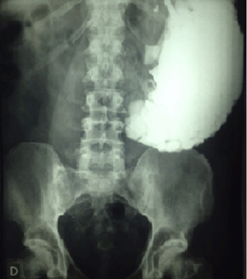
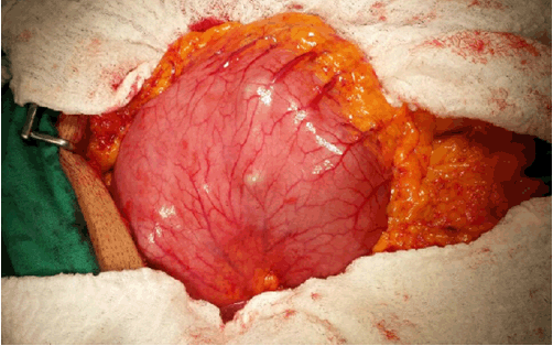
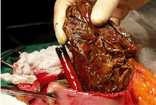
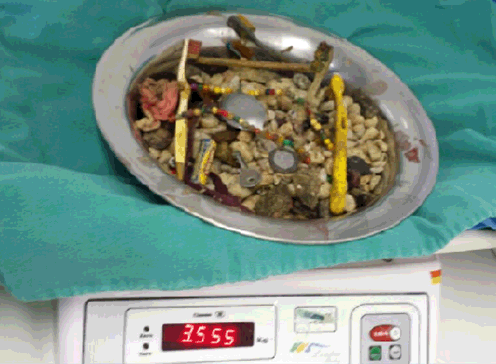
A 49-year-old male, schizophrenic patient, was presented to the emergency department. His relatives informed a progressive hyporexia over a four-month period associated with post-prandial fullness and a 10-kg weight loss. His past medical history was remarkable for metal pica (coins) two years before. Physical examination revealed mild pallor, epigastric bulging which was dull to percussion and an enormous hard, irregular, palpable mass, extending from the epigastrium to the left iliac fossa. Upon mobilization of the mass, a peculiar sound was produced, which was a clue for the diagnosis. Plain abdominal radiograph showed no signs of intestinal obstruction and a dilated stomach with a giant radiopaque bezoar containing different-sized grouped smaller images. This finding was another important evidence for the diagnosis (Figure 1). The patient was taken to open surgery, with evidence of a grossly dilated stomach (Figure 2) and performance of a longitudinal gastrotomy (Figure 3), manual removal of the foreign bodies (Figure 4) and a two-layer suture. At closer examination of the gastric mucosa, two superficial ulcers were identified, at the lesser and greater curvature, most likely due to contact with the sharp objects. There was neither any evidence of foreign bodies in other organs nor any complications. A 3.5-kg bezoar (Figure 5) comprised diverse objects: one plastic bag, two batteries, three lighters, six toothbrushes, some shoelaces, pens, coins, keys, spoons and necklaces, but predominantly stones in countless number. The patient's postoperative evolution was uneventful, with multidisciplinary care. Additionaly, he was cared for under close vigilance, with physical and chemical restraints to avoid pica. The diet was resumed in the third postoperative day and he was transferred on the seventh day to a referral psychiatric hospital. | ||||||
| ||||||
| ||||||
| ||||||
| ||||||
| ||||||
|
Discussion
| ||||||
|
Lithobezoars (stones) are quite rare, with very few cases described, as are metal bezoars and plastic bezoars [1][2] [4][5]. Most of the published data consist of case reports of colonic lithobezoars in children and adults [4] [6]. To the best of the our knowledge, there is only another article detailing a gastric lithobezoar which presented as dyspepsia, however, with much less stones compared to the current case [7]. Risk factors for bezoars in general include previous gastric surgery, conditions of reduced acidity or delayed motility, poor mastication, mental retardation, psychiatric disorders, and pica, i.e., an abnormal eating behavior characterized by ingestion of nonnutritive substances and classified according to the consumed matter: trichophagia (hair), acuphagia (sharp objects), lithophagia, etc. [3] [6][8]. In this case, the patient was schizophrenic and had previous metal pica. Usually, there are no symptoms until the bezoar reaches a substantial size but many patients remain oligosymptomatic. Gastric bezoars ordinarily cause dyspepsia (80%), distention, nausea, vomiting, hyporexia and halitosis. Complications as ulcers and perforation can occur [3] [5]. On physical examination, sometimes, it is possible to palpate the organ filled with the stones, with a characteristic "crunch sign", which usually applies to the colon [4] [9][10]. In the present case, given the palpation of the gastric bezoars with resulting singular sound, we strongly believe the concept also fits the stomach. Hence, we would like to propose the "gastric crunch sign". Radiographical diagnosis is especially valuable in litho bezoars, showing typically conglomerated radiopaque images within the organ. This unique appearance, "corn on the cob" sign, is considered pathognomonic. In this particular type of bezoar there is no need for barium studies or other image methods [4] [9] [10] . Upper endoscopy can be utilized in the diagnostic approach [3]. Nevertheless, the authors did not perform it given the large amount of foreign material in the stomach, making air insufflation particularly challenging, with risk of perforation. Gastrointestinal bezoars can be effectively treated by endoscopy or surgery. Endoscopic extraction is the choice in little or fragmentable bezoars, utilizing snares, rat-tooths, Dormia baskets and lithotripsy methods [1] [3][5]. Nevertheless, endoscopic management carries the risk of distal migration of fragments and iatrogenic injuries due to the manifold introductions/retrievals of the endoscope [3]. Open surgery is indicated in cases of failure of conservative attempts, large dimension bezoars composed of unfragmentable matter, complications such as intestinal obstruction, perforation and hemorrhage. The preferred approach is longitudinal gastrotomy in the anterior wall with removal of the bezoars followed by gastrorrhaphy [3]. We are successfully resorted to surgery and the patient had a favorable outcome. | ||||||
|
Conclusion
| ||||||
|
In conclusion, pica with resulting bezoar should be suspected in psychiatric patients presenting with nonspecific symptoms. Plain abdominal radiograph, a simple and widely available test, should be performed in those patients, confirming the diagnosis in case of radiopaque objects. Giant upper lithobezoars are very rare, can produce a gastric "crunch sign" and can be treated effectively by open surgery. | ||||||
|
References
| ||||||
| ||||||


|
[HTML Abstract]
[PDF Full Text]
|
|
Author Contributions
Alana Costa Borges – Substantial contributions to conception and design, Acquisition of data, Analysis and interpretation of data, Drafting the article, Revising it critically for important intellectual content, Final approval of the version to be published Marco Augusto Sobreira Rocha Filho – Analysis and interpretation of data, Revising it critically for important intellectual content, Final approval of the version to be published Vitor de Vasconcelos Muniz – Analysis and interpretation of data, Revising it critically for important intellectual content, Final approval of the version to be published |
|
Guarantor of submission
The corresponding author is the guarantor of submission. |
|
Source of support
None |
|
Conflict of interest
Authors declare no conflict of interest. |
|
Copyright
© 2016 Alana Costa Borges et al. This article is distributed under the terms of Creative Commons Attribution License which permits unrestricted use, distribution and reproduction in any medium provided the original author(s) and original publisher are properly credited. Please see the copyright policy on the journal website for more information. |
|
|