| |
|
|
|
Case Report
| ||||||
| Scalp pilonidal sinus: A case report | ||||||
| Salih AM1, Kakamad FH2 | ||||||
|
1Faculty of Medical Sciences, School of Medicine, Department Surgery, University of Sulaimani, François Mitterrand Street, Sulaymaniyah/IRAQ.
2Faculty of Medical Sciences, School of Medicine, Department Cardiothoracic and Vascular Surgery, University of Sulaimani, François Mitterrand Street, Sulaymaniyah, IRAQ. | ||||||
| ||||||
|
[HTML Abstract]
[PDF Full Text]
[Print This Article]
[Similar article in Pumed] [Similar article in Google Scholar] 
|
| How to cite this article |
| Salih AM, Kakamad FH. Scalp pilonidal sinus: A case report. Int J Case Rep Images 2016;7(3):175–177. |
|
Abstract
|
|
Introduction:
Pilonidal sinus is a chronic inflammatory condition which includes penetration of hair fragments into the skin. It rarely occurs in areas other than sacrococcygeal region.
Case Report: A 19-year-old male presented with pilonidal sinus of the posterior part left temporal area of the scalp for one year duration treated by complete excision and primary repair. Conclusion: Although extremely rare, pilonidal sinus may occur in the scalp with unknown etiology. | |
|
Keywords:
Hair containing sinus, Pilonidal sinus, Scalp, Sinus
| |
|
Introduction
| ||||||
|
Pilonidal disease is a common medical condition that accounts for almost 15% of anal suppurations [1]. It is a chronic inflammatory disease associated with the penetration of hair fragments into the skin. It occurs commonly in the sacrococcygeal region [2]. However, it may also occur in other areas rarely like umbilicus, nose, suprapubic area, axilla, groin, interdigital web, clitoris, prepuce, or penis [3]. The onset of pilonidal sinus is rare before puberty and after the age of forty [4]. It occurs three times more in male than female [4]. It clinically presents as mass, pain, cellulitis and redness [5]. Scalp pilonidal sinus is an extremely rare condition with only seven reported cases in literatures. Although trauma accounts for etiology of some cases, its exact cause remains unknown [2] [6] [7]. We present a case of scalp pilonidal sinus with literature review. | ||||||
|
Case Report
| ||||||
|
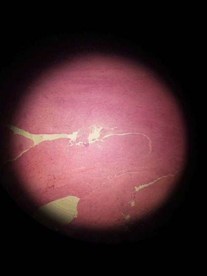
A 19-year-old male presented with a chronic nodule in the scalp for one year duration with intermittent discharge. There was no history of trauma. Local examination showed a single mobile nodule, 2x2 cm in size in the left parieto-occipital region with a punctum in center. Complete blood count, erythrocyte sedimentation rate and skull X-ray were normal. Complete excision of the nodule was performed under local anesthesia. Primary closure was done after irrigating the wound with diluted povidone. Histopathological examinations showed skin and subcutaneous tissue of scalp containing a sinus tract in the center infiltrated by mixed inflammatory cells, associated with foreign body giant cell reaction which contained hair particles. The wound showed clear margins after three months of the operation (Figure 1). | ||||||
| ||||||
| ||||||
|
Discussion
| ||||||
|
Pilonidal sinus refers to any subcutaneous sinus which contains hair [2]. It is a blind-end tract lined with granulation tissue, which leads to a cystic cavity lined with epithelial tissue [3]. The origin of pilonidal disease is not well understood. There are two theories associated with its pathogenesis: the acquired and the congenital theories. However, the majority of opinion favors the acquired theory [8] [9]. In general, at least three conditions need to be fulfilled for a pilonidal sinus to develop: First is hair in the skin and, second, some kind of wrinkled skin, such as the natal cleft or a scar. The third condition is a mixture of hormonal and hygienic problem [7]. It usually presents as pain, local inflammation and redness [5]. Treatment for symptomatic pilonidal sinus involves surgery to incise and drain the abscess. The surgery can be either wide excision and healing by secondary intention (longer healing time, low chance of recurrence), excision and primary closure by sutures (quicker healing, high risk of recurrence), or plastic surgical technique (for recurring and/or extensive sinus). The other procedures include topical application of natural polyphenols/laser epilation [3]. Scalp pilonidal sinus is a rare variant of pilonidal sinus. In most cases, its etiology remains unknown [2] [6] [7][10]. In some reported cases, trauma was accounted for the etiology by their authors [11] [12] [13]. Our patient did not report head trauma. Pilonidal sinus typically occurs in the sacrococcygeal area [2]. However, it may appear in other areas like axilla, groin, interdigital web, umbilicus, nose, suprapubic area, clitoris, prepuce, or penis [3]. Scalp pilonidal sinus is very rare. Table 1 gives scalp pilonidal sinus literature review with localization and proposed etiology. | ||||||
| ||||||
|
| ||||||
|
Conclusion
| ||||||
|
Although extremely rare, pilonidal sinus may occur in the scalp. Its etiology remains unknown. It can be excised and primarily sutured using local anesthesia. | ||||||
|
References
| ||||||
| ||||||
|
[HTML Abstract]
[PDF Full Text]
|
|
Author Contributions
Abdulwahid M. Salih – Substantial contributions to conception and design, Acquisition of data, Analysis and interpretation of data, Drafting the article, Revising it critically for important intellectual content, Final approval of the version to be published Fahmi H. Kakamad – Analysis and interpretation of data, Revising it critically for important intellectual content, Final approval of the version to be published |
|
Guarantor of submission
The corresponding author is the guarantor of submission. |
|
Source of support
None |
|
Conflict of interest
Authors declare no conflict of interest. |
|
Copyright
© 2016 Abdulwahid M. Salih et al. This article is distributed under the terms of Creative Commons Attribution License which permits unrestricted use, distribution and reproduction in any medium provided the original author(s) and original publisher are properly credited. Please see the copyright policy on the journal website for more information. |
|
|