| |
|
|
|
Case Report
| ||||||
| Subserosal fibroid thought to be recurrent granulosa cell tumor in a pregnant patient | ||||||
| Hiba J. Mustafa | ||||||
|
Queens Hospital Center, 82-68 164 Street, Jamaica, NY, 11432
| ||||||
| ||||||
|
[HTML Abstract]
[PDF Full Text]
[Print This Article]
[Similar article in Pumed] [Similar article in Google Scholar] 
|
| How to cite this article |
| Mustafa HJ. Subserosal fibroid thought to be recurrent granulosa cell tumor in a pregnant patient. Int J Case Rep Images 2016;7(3):165–169. |
|
Abstract
|
|
Introduction:
Juvenile androgen secreting granulosa cell tumors are very rare. We are describing a patient whom thought to have recurrence in pregnancy but turned to be subserosal pedunculated fibroid.
Case Report: We are presenting a case of a 30-year-old female with history of androgen secreting juvenile granulosa cell tumor of the ovary who underwent exploratory laparotomy for suspected recurrent granulosa cell tumor while she was 16 weeks pregnant. The mass turned out to be subserosal pedunculated fibroid. Conclusion: Recurrent juvenile androgen secreting granulosa cell tumor is a rare case. It can be confused with pedunculated fibroids even on MRI scan. Proceeding with laparoscopy first before exploratory laparotomy should be considered. | |
|
Keywords:
Androgen secreting type, Juvenile granules cell tumor, Uterine fibroid
| |
|
Introduction
|
|
Juvenile type granulosa cell tumors, defined in 1976 by Scully as a separate unit are found mostly in girls during the first two decades of life. On inspection under an optic microscope they are characterized by the presence of non-differentiated "blastemoid" structures and the formation of solid follicular formations and cysts. It comprises 5% of all granulosa cell tumors [1] [2]. This subtype tends to have a higher proliferative rate than the adult type and a lower risk for late recurrences. Granulosa cell tumors typically present as large masses; the mean diameter is 12 cm. Women may present with an asymptomatic mass noted on abdominal or pelvic examination. Often produce estrogen and/or progesterone; consequently, symptoms related to hyperestrogenism are common at diagnosis. Androgen production is rare and produces virilization in women [3] [4]. For women with stage I disease who wishes to preserve fertility or avoid exogenous hormone replacement, a unilateral salpingo-oophorectomy and uterine preservation with other procedures for complete surgical staging are appropriate [5]. Approximately, half of all pregnancy-associated stromal tumors are granulosa cell tumors, one-third are Sertoli-Leydig cell tumors, and the remainder are unclassified stromal tumors. The optimal time for semi-elective surgery during pregnancy is after the first trimester. In most cases, the preoperative workup for a pregnant woman with a pelvic mass can be limited to ultrasound imaging. If the ultrasound findings cannot distinguish between a possible pedunculated or degenerating leiomyoma and an ovarian neoplasm, obtaining magnetic resonance imaging (MRI) scan can be helpful. The more precise diagnosis afforded by MRI scan may allow the clinician to cancel planned exploratory surgery. |
|
Case Report
|
|
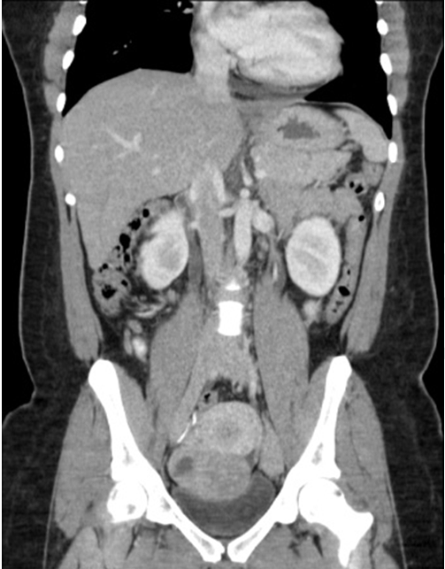
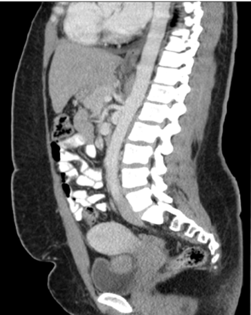
We present a case of 30-year-old Jamaican female who presented to gynecological oncology clinic in 2005 with hirsutism, clitoromegally of 3 cm and acne for one year. Workup showed elevated total testosterone to 365 ng/dl and 17-OH progesterone levels to 253 ng/dl. ACTH stimulation test was done to rule out late onset 21 hydroxylase deficiency which was ruled out. Pelvic Ultrasound revealed 11x7x10 cm left adnexal solid mass suspicious for tumor (Figure 1). Endometrial biopsy was done which showed benign endometrium and endocervical glands. Patient underwent exploratory laparotomy, right salpingo-oophorectomy, lymph nodes sampling, pelvic washings and partial omentectomy. Pathology results revealed androgen secreting granulosa cell tumor of the ovary stage IA. Testosterone levels normalized after the surgery. Patient underwent surveillance in gynecological oncology clinic with physical examinations, testosterone level, inhibin A and B levels and imaging, all were within normal limits. Patient followed with endocrine for continuous hirsutism and clitoromegally. Medical treatment of hirsutism was attempted with minimal response. In 2014, CT scan showed 7 cm adnexal mass with possible recurrence. Patient subsequently got pregnant (Figure 2) and (Figure 3). Testosterone and inhibin levels were still within normal limits. MRI scan showed 8x8x7 cm pelvic mass with cystic changes separate from the uterus (Figure 4) and (Figure 5). Case was discussed in tumor board with the presence of gynecological oncology, medical oncology, surgical oncology, radiology and MFM team. Anonymous decision was made to offer the patient surgery for suspected recurrent granulosa cell tumor of the ovary. Risks, benefits alternatives were explained to the patient. The team proceeded with exploratory laparotomy via vertical midline incision in which 8 cm lower uterine segment pedunculated Fibroid was noted and was excised. Results were confirmed by frozen section and later by pathology. Fetal monitoring was done during the procedure without fetal complications noted. |
|
|
|
|
|
|
|
|
|
|
|
Discussion
|
|
We are presenting a very rare case in multiple aspects: 1) Juvenile granulosa cell tumor: The juvenile type comprises 5% of all granulosa cell tumors, on histopathology has a macrofollicular or cystic pattern and is comprised of immature granulosa cells with frequent mitoses; Call-Exner bodies and coffee-bean grooved nuclei are not frequent. The histologic diagnosis is facilitated by immunohistochemical staining (IHC) using antibodies against markers of sex cord-stromal differentiation. Inhibin is the most sensitive and specific. Calretinin is typically positive, but is not specific for sex cord-stromal differentiation. Other markers, including CD99, Müllerian inhibiting substance, Vimentin, WT1, SF-1, cytokeratin, S-100 protein, and smooth muscle actin, are not specific and are not particularly helpful in distinguishing between granulosa cell tumor and its mimics. However, even positivity for inhibin is not absolutely specific for an ovarian sex cord tumor, as sex cord-stromal differentiation can be seen in other neoplasms [6]. 2) Androgen secreting granulosa cell tumor: Granulosa cell tumors often produce estrogen and/or progesterone; consequently, symptoms related to hyperestrogenism are common at diagnosis. Increased production of estrogen may also cause breast tenderness, postmenopausal bleeding, menstrual abnormalities, and, in children, sexual precocity. In our patient she presented with virilization due to the androgen secreting tumor which is very rare case scenario [7] [8]. 3) Persistence of the virilization after removal of the tumor. Our patient had persistent hirsutism and clitoromegally for many years after removal of the tumor in which hirsutism had almost no response to medical or mechanical therapy. Multiple types of imaging show possible recurrence of the tumor and now the patient is pregnant [9]. Ultrasound, CT scan, MRI scan all suggesting high possibility of tumor recurrence. Case was discussed in a multidisciplinary setting with anonymous decision to proceed with surgery in the second trimester for recurrent granulosa cell tumor of the ovary in which the mass appeared to be subserosal pedunculated fibroid [10] [11]. |
|
Conclusion
|
|
Juvenile type granulosa cell tumor is extremely rare cause of virilization. Persistence of virilization is also rare after removal of the tumor. Despite almost certainatity about a diagnosis in a pregnant patient we recommend considering laparoscopy before proceeding with exploratory laparotomy which could have been attempted in this patient as even though multiple images were obtained through ultrasound, CT scan and MRI the pedunculated fibroid was thought to be recurrent ovarian tumor. |
|
References
|
|
|
[HTML Abstract]
[PDF Full Text]
|
|
Author Contributions
Hiba J. Mustafa – Substantial contributions to conception and design, Acquisition of data, Analysis and interpretation of data, Drafting the article, Revising it critically for important intellectual content, Final approval of the version to be published |
|
Guarantor of submission
The corresponding author is the guarantor of submission. |
|
Source of support
None |
|
Conflict of interest
Authors declare no conflict of interest. |
|
Copyright
© 2016 Hiba J. Mustafa. This article is distributed under the terms of Creative Commons Attribution License which permits unrestricted use, distribution and reproduction in any medium provided the original author(s) and original publisher are properly credited. Please see the copyright policy on the journal website for more information. |
|
|