  |

|
 |
|
Clinical Image
| ||||||
| Left main coronary artery originating from the non-coronary sinus: A rare low-risk coronary anomaly | ||||||
| Razan F. Binyousef1, Fahed K. Habib2, Mohammed W. Althobaiti3 | ||||||
|
1MBBS, Radiologist, Medical Imaging Department, King Abdullah Medical City, Makkah, Saudi Arabia.
2MBBS, Radiologist, Medical Imaging Department, Alnoor Specialist Hospital, Makkah, Saudi Arabia. 3MD, Assistant professor, King Saud bin Abdulaziz university for health sciences KSAU-HS, consultant cardiac imaging, King Abdulaziz Medical City, Jeddah, Saudi Arabia. | ||||||
| ||||||
|
[HTML Abstract]
[PDF Full Text]
[Print This Article]
[Similar article in Pumed] [Similar article in Google Scholar] 
|


| How to cite this article |
| Binyousef RF, Habib FK, Althobaiti MW. Left main coronary artery originating from the non-coronary sinus: A rare low-risk coronary anomaly. Int J Case Rep Images 2016;7(3):195–197. |
|
Case Report
| ||||||
|
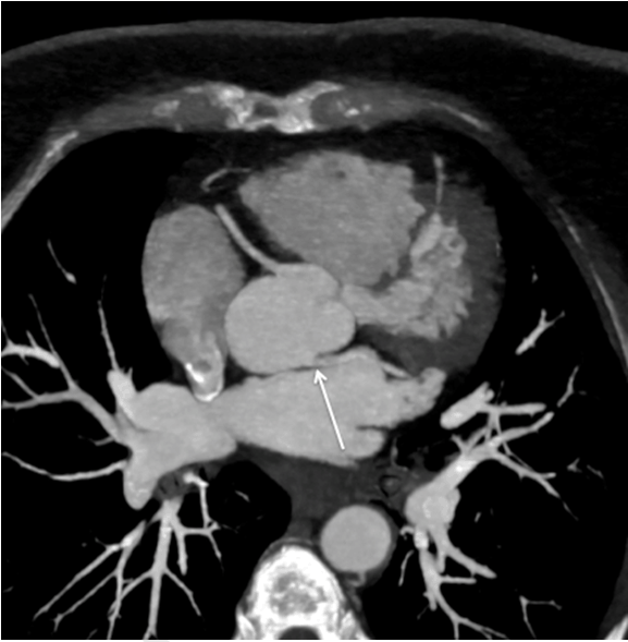
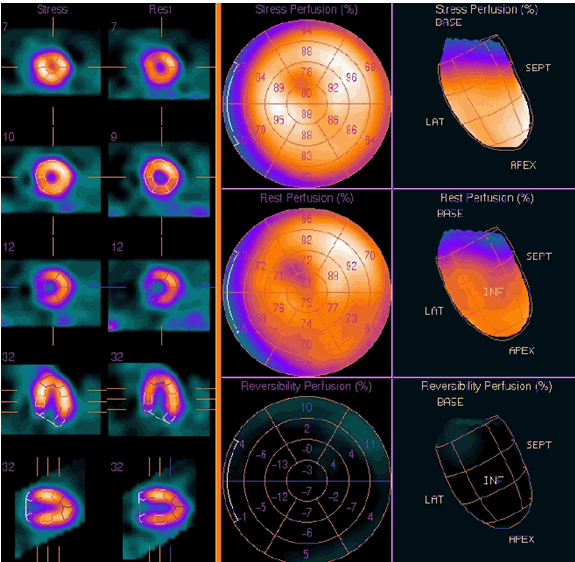
A 51-year-old female was evaluated for chest discomfort, palpitations, and worsening dyspnea on exertion. She had no relevant past medical history and the cardiac enzymes were within normal limits. She denied any history of sudden cardiac death in her family. On physical examination, she was noted to have a 1/6 systolic murmur heard over the second left inter-costal space and an electrocardiogram was normal. Transthoracic echocardiography was normal. A contrast enhanced cardiac computed tomography angiography (CTA) was performed to further evaluate her symptoms and to investigate for a possible coronary artery disease. The computed tomography scan showed the left main coronary artery arising from the non-coronary sinus with an acute angle (LCANCS) (Figure 1). A nuclear exercise stress test performed and was normal (Figure 2). | ||||||
|
| ||||||
|
| ||||||
| ||||||
|
Discussion
| ||||||
|
Anomalies of coronary arteries encompass a diverse group of rare coronary artery variations in origin, course or termination. Numerous studies reported incidence of coronary anomalies variably between 1% and 2% [1]. The reported incidence of coronary artery anomalies in patients undergoing coronary catheterization is 1.3% [1]. A retrospective series of 2096 patient undergoing coronary CTA showed a prevalence of approximately 2% [2]. The anomalous origin of left main coronary artery from non-coronary sinus is an extremely rare anomaly with variable incidence estimated at 0.0008% [1]. To the best of our knowledge, there are only three cases of LCANCS described by CT scan [3] [4] [5]. Anomalous origin of the left main coronary artery from the non-coronary sinus (LCANCS) is extremely rare with few reported cases in English literature [3]. A few reported cases of LCANCS were presented by acute myocardial infarction (MI), fatal arrhythmias and sudden cardiac death (SCD). Multiple studies proposed several mechanisms for high-risk coronary anomalies; these mechanisms include acute take-off angle (if the angle measures <45° between the proximal LCA and ascending aorta), intramural course (shared intima and media between aorta and LCA), or slit like ostium (if the ridge exceeded 50% of the coronary ostial luminal area) [6]. While the mechanisms are variable, the true significance of these coronary findings is not clear [7]. Not all coronary artery anomalies are felt to increase the risk of sudden cardiac death. The symptoms related to high risk coronary anomalies are mostly experienced during vigorous exercise. Most of the symptomatic cases are reported in young patients below the age of 30 [3]. Many cases presented late in relation to development of atherosclerotic changes. In elderly, these findings are probably detected incidentally in asymptomatic candidates. If these anomalies were associated with symptoms, the question would be what should be done next. Single-photon emission computed tomography (SPECT) myocardial perfusion test is a good option. A normal myocardial SPECT would obviate the need for further investigation particularly in old patients. However, if the SPECT myocardial perfusion test is positive in the presence of coronary anomaly in symptomatic patient, this would raise the question of whether surgical intervention should be expedited. In our case, the normal SPECT myocardial perfusion study was reassuring, excluding a significant coronary compression during stress and suggests that the anomaly in our patient is a benign incidental finding. | ||||||
|
Conclusion
| ||||||
|
In the present case, computed tomography angiography (CTA) identified a rare coronary anomaly and excluded the presence of obstructive atherosclerotic coronary artery disease substantiating its role as a powerful non- invasive imaging modality in the assessment of coronary arteries. Combining the functional data from nuclear stress test with anatomical data from cardiac CT scan excluded the presence of ischemia in the myocardial regions supplied by the anomalous coronary artery. Keywords: Anomalous left coronary artery, Coronary artery anomalies, Left main coronary artery from the non-coronary sinus (LCANCS) | ||||||
|
References
| ||||||
| ||||||

|
[HTML Abstract]
[PDF Full Text]
|
|
Author Contributions
Razan F. Binyousef – Substantial contributions to conception and design, Acquisition of data, Analysis and interpretation of data, Drafting the article, Revising it critically for important intellectual content, Final approval of the version to be published Fahed K. Habib – Substantial contributions to conception and design, Acquisition of data, Drafting the article, Final approval of the version to be published Mohammed W. Althobaiti – Substantial contribution to conception and design, Acquisition of data, Analysis and interpretation of data, Drafting the article, Critical revision of the article, Final approval of the version to be published |
|
Guarantor of submission
The corresponding author is the guarantor of submission. |
|
Source of support
None |
|
Conflict of interest
Authors declare no conflict of interest. |
|
Copyright
© 2016 Razan F. Binyousef et al. This article is distributed under the terms of Creative Commons Attribution License which permits unrestricted use, distribution and reproduction in any medium provided the original author(s) and original publisher are properly credited. Please see the copyright policy on the journal website for more information. |
|
|