| |
|
|
|
Case Report
| ||||||
| Multiple primary cancers in a patient and CT scan features of inferior vena cava leiomyosarcoma: A case report | ||||||
| Muhammad Imran Butt1, Muhammad Asif Shahzad2 | ||||||
|
1Department of Medicine Sir Charles Gairdner Hospital, Hospital Avenue, Nedlands, Perth WA 6009, Australia.
2Department of Medicine Lyell McEvin Hospital, Haydown Rd, Elizabeth Vale, SA 5112, Australia. | ||||||
| ||||||
|
[HTML Abstract]
[PDF Full Text]
[Print This Article]
[Similar article in Pumed] [Similar article in Google Scholar] 
|
| How to cite this article |
| Butt MI, Shahzad MA. Multiple primary cancers in a patient and CT scan features of inferior vena cava leiomyosarcoma: A case report. Int J Case Rep Images 2016;7(1):51–54. |
|
Abstract
|
|
Introduction:
Primary inferior vena cava (IVC) sarcoma is a rare tumor of mesenchymal origin arising from the tunica media of inferior vena cava. It accounts for about 0.5% of all soft tissue sarcomas and there are less than 300 cases reported in literature [1][2]. It is the most common vascular sarcoma [3]. To the best of our knowledge, it has not been reported in combination with any other primary malignancy.
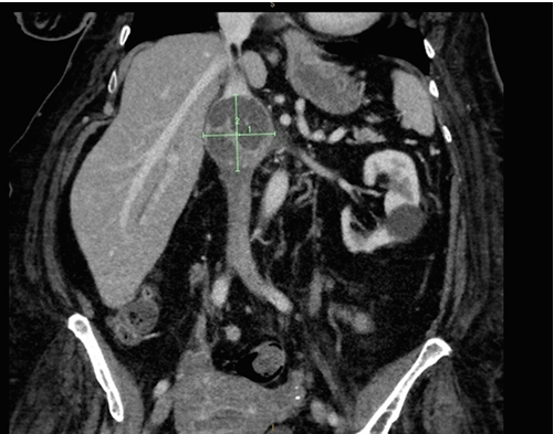
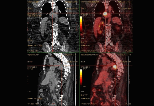
Case Report: An 86-year-old female with breast cancer presented with progressive development of bilateral lower limbs edema which was attributed to a palpable abdominal mass. Computed tomography scan revealed a large mass in her inferior vena cava (IVC) with features consistent with an IVC sarcoma. On PET scan, there was moderate grade heterogeneous activity related to the IVC mass, again suggestive of IVC sarcoma. Also, there was an intense uptake at gastroesophageal junction which was proven to be esophageal carcinoma on histology. Patient did not consent for biopsy of IVC lesion due to the risk involved in the procedure hence treated conservatively for edema with moderate improvement and received radiotherapy for esophageal carcinoma with good tolerance. Conclusion: Multiple malignancies are uncommon and a long-term meticulous follow is required to diagnose second malignancy at early stage to reduce mortality rate. To our knowledge, this is the first ever case of this combination of primary malignancies with IVC sarcoma being a rare tumor. | |
|
Keywords:
Inferior vena cava, Leiomyosarcoma, Multiple Primary cancers
| |
|
Introduction
| ||||||
|
In last three decades, there have been dramatic improvements in cancer survival, mainly due to advances in treatment and the increased detection of cancer at an early stage. However, incidence of multiple primary malignancies has increased in recent decades. More recently in clinical trials and case control studies have shown a better understanding of risk factors for the development of multiple primary cancers. Cancer patients have a 20% greater risk of a new primary cancer compared with the general population. Also, second cancers have become a leading cause of death among long-term cancer survivors. The occurrence of a second cancer can be expected but third or higher order malignancies are rare [4]. | ||||||
|
Case Report
| ||||||
|
An 86-year-old female presented with progressive development of bilateral lower limbs pitting edema along with gradual increase in her weight from 85 kg to 102 kg over 6 months. This was associated with mild shortness of breath on exertion developing over one year. She did not report any chest pain, orthopnea or paroxysmal nocturnal dyspnea. She had past history of breast cancer treated with left sided mastectomy and left axillary lymph nodes (LNs) clearance along with postoperative radiotherapy. Ca Breast was 50 mm mass, grade 1, estrogen receptor (ER) positive and HER2 (human epidermal growth factor receptor 2) negative invasive ductal cell carcinoma with 12 positive axillary LNs. She was also commenced on adjuvant therapy with Tamoxifen. Her other medical history included type 2 diabetes mellitus, hypertension and Barrett's oesophagus. Her medications included amlodipine, pioglitazone, metformin, and tamoxifen. On examination, her vital signs were within normal limits. Cardiovascular and respiratory examinations were unremarkable. However, a mass was palpable in epigastrium on deep palpation. Her ECG showed normal sinus rhythm with no signs of right or left ventricular hypertrophy. This was subsequently confirmed by echocardiogram with no significant structural abnormality. Computed tomography scan of abdomen revealed a large 56x53 mm heterogeneous mass within the IVC with imperceptible caval lumen and extended into the orifice of the left renal vein (Figure 1). Similar changes were noted where the right renal vein inserts into the IVC. Overall appearance favored an IVC sarcoma. There were no mesenteric or retroperitoneal nodes enlargement and there was no evidence for bone metastasis. A PET scan was done which showed moderate grade heterogeneous activity associated with the fusiform mass in IVC again suggestive of sarcoma. Also, there was an intense activity at gastroesophageal junction highly suspicious for a primary tumor (Figure 2). On further investigations, gastroscopy showed large Hiatus hernia with moderate sized semi circumferential mass on cardia. The mass was not obstructive and was extending into distal esophagus for 5–7 mm. Esophageal biopsy proved an invasive moderately differentiated adenocarcinoma characterised by irregular malignant glands within fibrotic stroma. Patient did not provide consent to have biopsy of IVC lesion due to the risk involved in the procedure and opted for conservative management with frusemide with some improvement in her symptoms. Oncology and radiotherapy opinions were sought and she was deemed not suitable for aggressive chemotherapy for either of the lesions at that stage, however, received low dose radiotherapy for esophageal cancer with reasonable tolerance. | ||||||
| ||||||
| ||||||
|
Discussion
| ||||||
|
The exact incidence of triple tumors in one patient remains unknown [5]. A literature review on 1,104,269 cancer patients concluded that the prevalence of multiple primary malignancies occurs in 11.7% [6]. In another study encountered in 3–5% of malignant tumors, triple tumors occur in only 0.5%, quadruple tumors in 0.3% of malignant tumors [7]. Risk factors for multiple primary tumors include genetic, constitutional and environmental factors. None were detected in our patient. Unfortunately, our patient did not undergo detailed genetic testing to uncover possible underlying genetic mutations. Malignant synchronous tumors (i.e., diagnosed simultaneously or with two months of each other) are predominantly seen in oropharyngeal area, gastrointestinal tract, lungs, breasts, urinary tract and genital organs. Usually, synchronous tumors have same histological features. Metachronous cancers (diagnosed in time interval >2 months) in addition to above mentioned locations are more frequently lymphoma and myeloma in patients with gastric cancer; colorectal cancer in patients with endometrial cancer and thyroid cancer in patients with respiratory system cancers [8]. The set of malignant tumors in our patient is unusual and does not fit any of the multiple primary cancer syndromes described thus so far. Between January 1993 and September 2006, of 1429 patients registered to a Sarcoma Center, IVC sarcoma was seen only in one patient [9]. It is an uncommon tumor with poor prognosis, however, with an aggressive surgical approach in association with the absence of metastasis a long-term survival or even cure can be expected [10] [11]. Tumor size is one of the main prognostic factors and five-year survival is observed in 30–53% of patients who undergo resection with free margins. It may be difficult to differentiate between a sarcoma of IVC and another soft tissue sarcoma of the retroperitoneum involving the IVC. The true sarcoma of IVC originates from smooth muscle cells of the vessel and its growth pattern may be either intra- or extraluminal, with possibility of invasion of adjacent structures [10]. According to the site involved IVC sarcomas are divided into 3 levels. Level 1 extends from the entry of the hepatic veins up to the right atrium. Level 2 comprises the area between the confluences of the renal and hepatic veins and, Level 3 is distal to the entry of renal veins to IVC [12] [13]. Non-invasive imaging modalities such as computed tomography scan, ultrasonography and magnetic resonance imaging scan are often used to diagnose these tumors. They provide valuable information regarding their origin, evaluating the presence of local invasion and excluding distant metastases. The sensitivity and specificity of CT in assessment of the tumors are 78% and 96% respectively and even higher with MRI (95–100%) [13]. Selective arteriography of the celiac trunk may also be performed in patients where hepatic invasion or metastasis is suspected and transesophageal echocardiography can exclude or verify intracardiac tumor extension [14]. Computed tomography scan typically reveal a retroperitoneal, lobulated, non-calcified mass, either with or without areas of low central attenuation resulting from necrosis or cystic degeneration, with no fat content and heterogeneous contrast enhancement [15]. Despite advancing imaging technology, a biopsy is still required for formal diagnosis. Histopathology of IVC sarcoma reveals spindle tumor cells, which are positive for markers of smooth muscle activity including vimentin, muscle actin, alpha-smooth muscle actin, and desmin [16]. In one study, it was found that the most useful sign for identifying a retroperitoneal mass as an IVC sarcoma was an imperceptible caval lumen. This was seen in 75% of sarcomas arising from the IVC but not in any case with an alternate diagnosis . This sign had both high positive and negative predictive values of 100% and 92% respectively. A negative embedded IVC sign is considered present if the IVC is displaced to the periphery of the mass, taking on a crescentic configuration which excludes IVC origin of the mass [11]. Prognosis of IVC Sarcoma | ||||||
|
Conclusion
| ||||||
|
Multiple malignancies occur rarely. The etiology remains controversial and a large number of cancer patients have to be followed for long periods to obtain adequate data about the development of subsequent additional malignancies. To our knowledge, this is the first detected case with this combination of primary cancers. An imperceptible inferior vena cava (IVC) at the point of maximal contact with a retroperitoneal mass was the most useful CT feature for predicting the origin of IVC sarcoma. A negative embedded organ sign was useful for excluding IVC origin. Knowledge of these CT features may assist with preoperative planning. | ||||||
|
References
| ||||||
| ||||||
|
[HTML Abstract]
[PDF Full Text]
|
|
Author Contributions
Muhammad Imran Butt – Substantial contributions to conception and study design, Analysis and interpretation of data, Revising it critically for important intellectual content, Final approval of the version to be published Muhammad Asif Shahzad – Acquisition of data, Drafting the article, Final approval of the version to be published |
|
Guarantor of submission
The corresponding author is the guarantor of submission. |
|
Source of support
None |
|
Conflict of interest
Authors declare no conflict of interest. |
|
Copyright
© 2016 Muhammad Imran Butt et al. This article is distributed under the terms of Creative Commons Attribution License which permits unrestricted use, distribution and reproduction in any medium provided the original author(s) and original publisher are properly credited. Please see the copyright policy on the journal website for more information. |
|
|