| |
|
|
|
Case Report
| ||||||
| A case of pericardial angiosarcoma with refractory pericardial tamponade treated with multidisciplinary therapy with pericardial fenestration, radiotherapy and chemotherapy | ||||||
| Hirano Satoshi1, Yamanaka Kyoko2, Ichinose Shuji3, Ikeda Atsushi4, Hayama Noriko5, Shimizu Shinichiro6, Aruga Takashi7, Uchida Osamu8, Nakamura Sukeyuki9 | ||||||
|
1Manager, Department of Medical Oncology, Funabashi Municipal Medical Center, Funabashi, Chiba, Japan.
2Senior resident, Division of Cardiology, Funabashi Municipal Medical Center Heart and Vascular Institute, Funabashi, Chiba, Japan. 3Assistant manager, Department of Thoracic Surgery, Funabashi Municipal Medical Center, Funabashi, Chiba, Japan. 4Assistant manager, Division of Cardiology, Funabashi Municipal Medical Center Heart and Vascular Institute, Funabashi, Chiba, Japan. 5Chief physician, Department of Respiratory Medicine, Funabashi Municipal Medical Center, Funabashi, Chiba, Japan. 6Manager, Laboratory Division of Pathology, Funabashi Municipal Medical Center, Funabashi, Chiba, Japan. 7Manager, Department of Radiation Oncology, Funabashi Municipal Medical Center, Funabashi, Chiba, Japan. 8Manager, Department of Thoracic Surgery, Funabashi Municipal Medical Center, Funabashi, Chiba, Japan. 9Manager, Department of Respiratory Medicine, Funabashi Municipal Medical Center, Funabashi, Chiba, Japan. | ||||||
| ||||||
|
[HTML Abstract]
[PDF Full Text]
[Print This Article]
[Similar article in Pumed] [Similar article in Google Scholar] 
|
| How to cite this article |
| Satoshi H, Kyoko Y, Shuji I, Atsushi I, Noriko H, Shinichiro S, Takashi A, Osamu U, Sukeyuki N. A case of pericardial angiosarcoma with refractory pericardial tamponade treated with multidisciplinary therapy with pericardial fenestration, radiotherapy and chemotherapy. Int J Case Rep Images 2015;6(11):707–711. |
|
Abstract
|
|
Introduction:
Outcomes for patients with pericardial angiosarcoma, especially with metastatic disease are very poor, even when these patients are treated with chemotherapy or radiotherapy.
Case Report: This report describes a case of a 50-year-old male with pericardial angiosarcoma presenting with cardiac tamponade. Repeat pericardiocentesis showed bloody fluid with cytopathology negative for malignant cells. Pericardial fenestration was performed to prevent recurrent pericardial tamponade due to poor drainage. A diagnosis of cardiac angiosarcoma was made based on the resected pericardium and a biopsied specimen from gluteus medius muscle. The patient responded to combination therapy with docetaxel and radiotherapy. However, the patient died four months after diagnosis due to intraperitoneal bleeding from liver metastases. The patient was not dependent on a chest tube or a pericardial drain before he died. At autopsy, only a small amount of residual tumor was revealed around the heart. Conclusion: There is a possibility that palliative local control against refractory cardiac tamponade could be obtained via multidisciplinary therapy with pericardial fenestration, radiotherapy, and chemotherapy. | |
|
Keywords:
Cardiac tamponade, Multidisciplinary therapy, Pericardial angiosarcoma, Pericardial fenestration, Radiotherapy
| |
|
Introduction
|
|
Pericardial angiosarcoma is extremely rare, but this entity should still be included in the differential diagnosis of bloody pericardial effusion, even when cytopathology is negative for malignant cells. Outcomes for patients with metastatic disease are very poor, even when these patients are treated with chemotherapy or radiotherapy. This report describes a case of a patient with pericardial angiosarcoma presenting with recurrent pericardial tamponade and illustrates that palliative local control against cardiac tamponade can be obtained via multidisciplinary therapy with pericardial fenestration, radiotherapy, and chemotherapy. |
|
Case Report
|
|
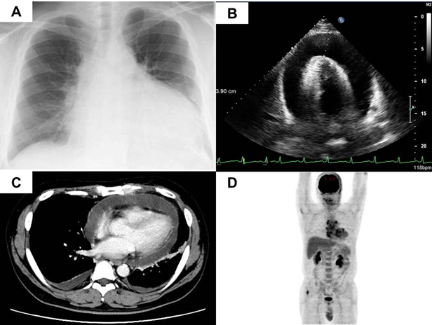
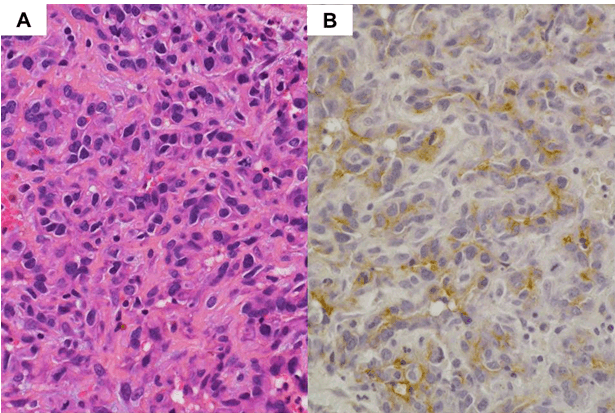
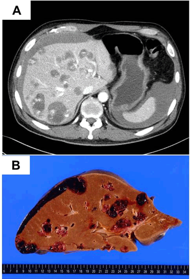
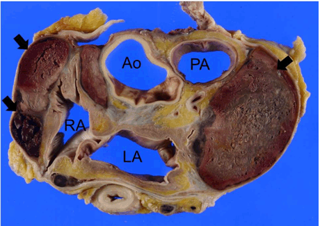
A 50-year-old male with a history of gout treated with febuxostat was referred for the evaluation for a three-day history of dyspnea and pericardial effusion. He admitted to heavy alcohol consumption and 28 pack-years of former smoking. On physical examination, his pulse was 108 bpm, blood pressure was 128/ 88 mmHg, and oxygen saturation was 98% on room air. His body temperature was 37.4°C. Heart murmurs and lung rales were not heard. The rest of the physical examination was unremarkable. His basic laboratory findings were unremarkable except for elevation of hepatocellular enzymes (aspartate aminotransferase of 42 IU/L and alanine aminotransferase of 50 IU/L) and C-reactive protein (3.03 mg/dL). Electrocardiogram showed sinus rhythm and low voltage tracings. Chest X-ray showed cardiac enlargement (Figure 1A). Echocardiography demonstrated massive pericardial effusion (Figure 1B). Pericardiocentesis was performed, and 1600 mL of bloody fluid was aspirated. Repeat fluid cytologic examination revealed no malignant cells. The results of examinations for tuberculosis and other bacterial agents were negative. A pericardial drain was placed, and drain output was 600 to 800 mL of bloody fluid every other day. Contrast-enhanced computed tomography (CT) scan of the chest showed large pericardial effusion and swelling of mediastinal lymph nodes (Figure 1C). There was no evidence of intracardiac abnormality. 18F-labeled deoxyglucose (FDG) positron emission tomography scan showed increased FDG uptake in mediastinum nodules, pericardium, gluteus medius muscle, and cervical vertebrate (Figure 1D). About two weeks later, the pericardial drain was removed to prevent infection. However, two weeks after removal of pericardial drain, the patient developed syncope with massive pericardial effusion. A pericardial drain was placed but was not effective due to the high viscosity of the pericardial fluid. Therefore, pericardial fenestration was performed to prevent recurrent cardiac tamponade. From the resected pericardium and the specimen from a gluteus medius muscle biopsy, a diagnosis of pericardial angiosarcoma was made. Proliferation of atypical short spindle or oval or epithelioid cells with hyperchromatic irregular nuclei and eosinophilic cytoplasm arranged in papillary structures or situated around variably gaping organization was demonstrated. Immunohistochemically, many atypical cells were positive for CD31, CD34, and Fli-1 (Figure 2). Desmin, S-100, D2-40, cytokeratins (CAM5.2, AE1/AE3), EMA, calretinin, and STAT6 were negative. The patient was treated with concomitant chemoradiotherapy using a total dose of 40 Gy (2 Gy/fraction) for 4 weeks and docetaxel at a dose of 25 mg/m2 weekly after administration of 300 mg of carboplatin into the pericardial space. The pericardial drain was removed after administration of carboplatin, and the chest drainage tube was also removed two weeks later. After 1 month, multiple brain, pulmonary, and liver metastases were demonstrated. Therefore, treatment with docetaxel was stopped, and pazopanib was initiated. Two months later, he presented to our hospital with palpitations, dyspnea on exertion, and new-onset dizziness. Laboratory work was performed and showed severe anemia with a hemoglobin of 5.6 g/dL. He received 2 units of red blood cells, but his condition did not improve. Two days later, he was readmitted to our hospital for further evaluation. His heart rate was 101 bpm, blood pressure was 93/65 mmHg, and oxygen saturation was 96% on room air. On initial laboratory evaluation, his hemoglobin level was 5.6 g/dL. He also complained of right upper abdominal pain. Contrast-enhanced CT scan of the abdomen showed intraperitoneal hemorrhage due to multiple liver metastases (Figure 3A). Unfortunately, the patient died four days after readmission due to intraperitoneal bleeding. At autopsy, fatal massive intraperitoneal hemorrhage arising from liver metastases was demonstrated (Figure 3B). Although multiple nodular tumors remained within the pericardium, only a small pericardial effusion was demonstrated (Figure 4). Microscopically, most of the pericardial tumors were necrotic. Multiple lung metastases were also demonstrated, but pleural effusion was mild (about 300 mL in each thoracic cavity). |
|
|
|
|
|
|
|
|
|
Discussion
|
|
Primary cardiac angiosarcoma is extremely rare but is the most common primary cardiac malignancy. Therefore, cardiac angiosarcoma should be included in the differential diagnosis of cardiac tamponade. Unfortunately, this tumor is not easy to diagnosis due to its minimal incidence and difficult location to approach. Pericardiocentesis and tissue biopsy are often used for diagnosis. However, pericardial fluid cytology analysis is unreliable and should not be used, because malignant cells are very rarely found in the bloody fluid, even when the tumor has invaded into the pericardium, as in the present case. For accurate diagnosis, surgical exploration and intraoperative frozen sections are needed [1]. Outcomes associated with these tumors are very poor due to their aggressive character and due to late diagnosis, with 80% of patients already presenting with metastases at the time of diagnosis [2]. Surgical resection of primary tumor should be attempted when feasible, as overall survival (OS) may be improved. Few studies have characterized survival outcomes in patients with cardiac angiosarcoma and metastatic disease. Hong et al. analyzed 10 patients who presented with metastatic disease and reported a median OS of only six months [3]. The optimal chemotherapy regimen for these aggressive tumors has not been established. Recently, Okiyama et al. reported that low-dose docetaxel therapy was effective for treatment of angiosarcoma of the skin [4]. On the other hand, Nakamura-Horigome et al. reported that docetaxel and radiotherapy was an effective treatment for cardiac angiosarcoma [5]. Their patient was treated with standard fractionated radiotherapy with a total dose of 42 Gy (2 Gy/fraction) for 4 weeks. The patient received docetaxel, 25 mg/m2 weekly. With this treatment, the radiation dosage was reduced, using docetaxel as a radiosensitizer to decrease the adverse effects of high-dose standard fractionated radiotherapy. Subsequent cycles docetaxel were administered as maintenance therapy (2 weeks of treatment, 1 week of rest) until disease progression. Suderman et al. described a case of cardiac angiosarcoma treated with the same method. Their patient survived for more than 16 months [6]. Pazopanib is a synthetic imidazolyl pyrimidine that functions as a multi-targeted tyrosine kinase inhibitor with a high affinity for vascular endothelial growth factor receptors. The median length of progression-free survival and overall survival of patients with angiosarcoma treated with pazopanib after one or more cytotoxic regimens was reported as 3.2 months and 8.0 months, respectively [7]. A recent case report noted a durable response of more than 10 months when using pazopanib for treatment of pericardial angiosarcoma [8]. In our patient, multiple metastases were demonstrated at the time of diagnosis, and recurrent pericardial effusion was hard to resolve. Therefore, multidisciplinary therapy with pericardial fenestration, radiotherapy, and chemotherapy with docetaxel followed by pazopanib was performed. The treatment was effective for local tumor control and for maintenance of quality of life. The patient was not dependent on a chest tube or a pericardial drain until the time of his death. |
|
Conclusion
|
|
We conclude that multidisciplinary approaches involving palliative surgery, radiotherapy, and chemotherapy may offer better quality of life in cases of cardiac angiosarcoma even in patients with metastatic disease. |
|
Acknowledgements
|
|
We thank Dr. Hisaoka Masanori for his diagnostic expertise. |
|
References
|
|
|
[HTML Abstract]
[PDF Full Text]
|
|
Author Contributions
Satoshi Hirano – Substantial contributions to conception and design, Acquisition of data, Analysis and interpretation of data, Drafting the article, Revising it critically for important intellectual content, Final approval of the version to be published Kyoko Yamanaka – Analysis and interpretation of data, Revising it critically for important intellectual content, Final approval of the version to be published Shuji Ichinose – Analysis and interpretation of data, Revising it critically for important intellectual content, Final approval of the version to be published Atsushi Ikeda – Analysis and interpretation of data, Revising it critically for important intellectual content, Final approval of the version to be published Noriko Hayama – Analysis and interpretation of data, Revising it critically for important intellectual content, Final approval of the version to be published Shinichiro Shimizu – Analysis and interpretation of data, Revising it critically for important intellectual content, Final approval of the version to be published Takashi Aruga – Analysis and interpretation of data, Revising it critically for important intellectual content, Final approval of the version to be published Osamu Uchida – Analysis and interpretation of data, Revising it critically for important intellectual content, Final approval of the version to be published Sukeyuki Nakamura – Analysis and interpretation of data, Revising it critically for important intellectual content, Final approval of the version to be published |
|
Guarantor of submission
The corresponding author is the guarantor of submission. |
|
Source of support
None |
|
Conflict of interest
Authors declare no conflict of interest. |
|
Copyright
© 2015 Satoshi Hirano et al. This article is distributed under the terms of Creative Commons Attribution License which permits unrestricted use, distribution and reproduction in any medium provided the original author(s) and original publisher are properly credited. Please see the copyright policy on the journal website for more information. |
|
|