  |

|
 |
|
Case Report
| ||||||
| Cervical paraspinal chordoma with left vertebral artery encasement | ||||||
| Chi-Man Yip1, Ping-Hong Lai2, Hui-Hwa Tseng3, Shu-Shong Hsu1 | ||||||
|
1Division of Neurosurgery, Department of Surgery, Kaohsiung Veterans General Hospital, Kaohsiung, Taiwan.
2Department of Radiology, Section of Neuroradiology Kaohsiung Veterans General Hospital, Kaohsiung, Taiwan. 3Department of Pathology, Kaohsiung Veterans General Hospital, Kaohsiung, Taiwan. | ||||||
| ||||||
|
[HTML Abstract]
[PDF Full Text]
[Print This Article]
[Similar article in Pumed] [Similar article in Google Scholar] 
|
| How to cite this article |
| Chi-Man Y, Ping-Hong L, Hui-Hwa T, Shu-Shong H. Cervical paraspinal chordoma with left vertebral artery encasement. Int J Case Rep Images 2015;6(11):698–701. |
|
Abstract
|
|
Introduction:
Chordomas are slow-growing, low-grade malignant but locally invasive tumors which originate from embryonic remnants of the primitive notochord. Chordomas are principally midline tumors. In the neuraxis, chordomas are most commonly located in the sacrococcygeal region (50–55%), followed by the cranio-occipital region (25–30%).
Case Report: A 71-year-old male has a left paraspinal tumor extending from C2 to C6 with bone erosion and left vertebral artery encasement. The tentative diagnosis before surgery was lymphoma or metastatic tumor. He underwent posterior cervical decompression with surgical debulking of the tumor to release the cord compression and posterior lamina screw fixation from C2 to C7 with allograft fusion and pathology confirmed the tumor to be chordoma. Conclusion: Due to the rare occurrence of chordomas extra-axially, these lesions have not earned a great deal of consideration in the clinical and radiographic differential diagnoses of extra-axial paraspinal lesions. An accurate preoperative diagnosis of chordoma is crucial, as survival is optimal when radical en bloc resection is performed at primary surgery. | |
|
Keywords:
Cervical paraspinal, Chordoma, Extra-axial paraspinal lesions, Radical en bloc resection
| |
|
Introduction
| ||||||
|
Chordomas are slow-growing, low-grade malignant but locally invasive tumors which originate from embryonic remnants of the primitive notochord. These tumors account for approximately 1% of all intracranial tumors, 4% of primary bone tumors and 2% of spinal tumors [1] [2] [3]. Chordomas are principally midline tumors. In the neuraxis, chordomas are most commonly located in the sacrococcygeal region (50–55%), followed by the cranio-occipital region (25–30%) and then in the mobile spine vertebral bodies (10–15%) [1] [3] [4] [5] [6]. For extra-axial paraspinal lesions, the common pathology are metastatic tumor, neurogenic tumor (schwannoma or neurofibroma) and lymphoma [2]. Extra-axial chordomas are rare but important consideration in the diagnosis of extra-axial lesions of the central nervous system. We would like to share a case of cervical paraspinal chordoma with others in order to increase clinical experience. | ||||||
|
Case Report
| ||||||
|
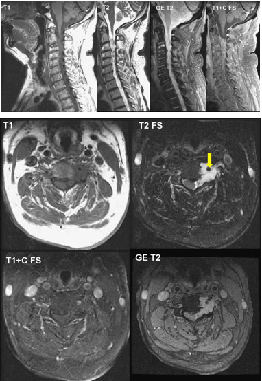
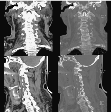
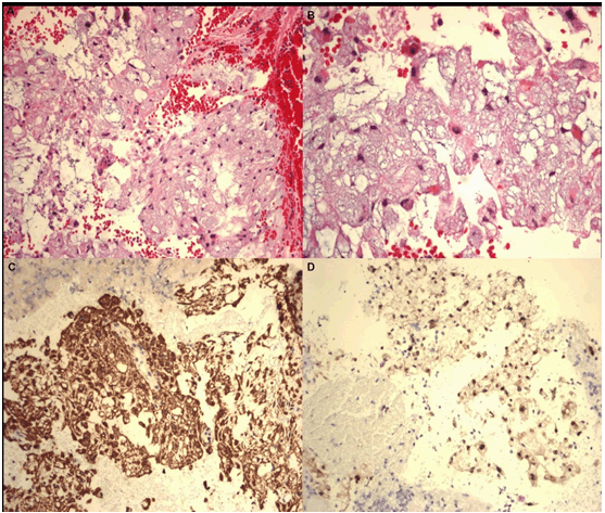
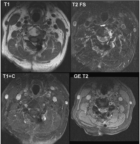
A 71-year-old male having past medical history of hypertension, herb-related chronic renal insufficiency and Parkinsonism who presented to us in December 2014 with the chief complains of progressive weakness of four extremities and unsteady gait for more than one year. On admission, he was conscious clear and his neurological examination revealed motor weakness of his bilateral upper limbs and bilateral lower limbs with muscle power grade 4; clumsy hands and unsteady gait; positive Hoffmann's sign and Babinski's sign on his left side; fine hand tremor; evident spasticity on his four limbs and rigidity on his upper limbs. C-spine plain film showed degenerative change, but magnetic resonance imaging (MRI) scan of C-spine (Figure 1A) (Figure 1B) demonstrated a mildly enhancing mass lesion involving left side spinal canal from C2 to C6, left neuroforamina of C2/3, C3/4, C4/5, C5/6, erosion of vertebral body, left pedicle, lamina and transverse foramina of C3 to C5, encasement of left vertebral artery (yellow arrow), and spinal cord compression at C3 level. Computed tomography (CT) scan of C-spine (Figure 2) showed multiple foci of radiolucent and osteolytic lesions over left lateral mass of C3, C4 and C5, as well as narrowing was found over left C3/C4, C4/C5 and C5/C6 neuroforamina. Based on the imaging finding, lymphoma or metastatic tumor was the tentative diagnosis, but the tumor markers checked before operation were within normal limits. Under general anesthesia, this patient was put in prone position with his neck was in neutral position. He underwent posterior cervical decompression with surgical debulking of the tumor to release the cord compression and posterior lamina screw fixation from C2 to C7 with allograft fusion. Histology examination of the specimen (Figure 3) revealed individual cells, cords and lobules of physaliferous cells and cells with eosinophilic cytoplasm in a prominent myxoid background. Nucleoli was inconspicuous and mitoses was absent. The immunohistochemistry results of neoplastic cells reveal positive for brachyury, EMA, S100, CKAE1/AE3, and negative for GFAP immunohistochemistry. Chordoma was diagnosed based on the tumor cells morphology and immunohistochemistry results. Followed-up C-spine MRI scan (Figure 4) showed that the spinal cord was free from the compression, however, residual tumor was present. Postoperative radiotherapy was arranged and the dosage was 45 Gy on the tumor bed in 25 fractions. He recovered well from the surgery and postoperative radiotherapy. He is regularly follow-up at our out-patient department in a stable condition. | ||||||
|
| ||||||
|
| ||||||
|
| ||||||
| ||||||
|
Discussion
| ||||||
|
Notochord is an embryonic structure which subsequently develops as vertebral column. During the second month of embryonic development, the notochord is restricted to the intervertebral residues. In adults, it gives rise to the nucleus pulposus of the intervertebral disks. Remnants of the notochord may persisted and give rise to a chordoma which can occur at any level along the neural axis [4] [7]. The radiological features of chordoma on MRI scan include hypointense or isointense on T1-weighted image (T1WI), hyperintense on T2-weighted image (T2WI), with heterogeneous enhancement. Low signal intensity fibrous septae on T2WI is detected in 70% cases [3] [4][5] [7]. However, these MRI findings are very nonspecific and the differential diagnosis includes neurofibroma, metastasis, lymphoma, and chondrosarcoma [7]. Computed tomography (CT) scan is helpful to assess the degree of bone involvement or destruction and detect patterns of calcifications within the lesion in 30–70% of cases. Imaging differential diagnosis of spinal extradural and foraminal/extraforaminal mass lesions includes neurogenic tumor (schwannoma, neurofibroma), metastasis, lymphoma and chordoma which is a very rare entity [2]. An accurate preoperative diagnosis of chordoma is crucial, as survival is optimal when radical en bloc resection is performed at primary surgery if possible [5] [8]. Spinal extraosseous chordoma arises from ectopic notochordal rests outside the vertebrae and it should be in the differential diagnosis when a soft tissue mass with dumbbell morphology, vertebral artery encasement, vertebral body bone erosion, high signal on T2WI and varied enhancement. Nuclear staining for brachyury represents a unique specific diagnostic marker for chordoma [9]. Aggressive resection is the treatment of choice, followed by adjuvant treatments (radiation and sometimes chemotherapy). Proton beam radiation has been used to treat chordomas recently [8]. | ||||||
|
Conclusion
| ||||||
|
Spinal extraosseous chordoma should be in the differential diagnosis when a soft tissue mass showing dumbbell morphology, vertebral artery encasement, vertebral body bone erosion, and hyperintense on T2WI image with varied enhancement. An accurate preoperative diagnosis of chordoma is crucial, as survival is optimal when radical en bloc resection is performed at primary surgery. | ||||||
|
References
| ||||||
| ||||||

|
[HTML Abstract]
[PDF Full Text]
|
|
Author Contributions
Chi-Man Yip – Substantial contributions to conception and design, Acquisition of data, Drafting the article, Revising it critically for important intellectual content, Final approval of the version to be published Ping-Hong Lai – Substantial contributions to conception and design, Analysis and interpretation of data, Revising the article critically for important intellectual content, Final approval of the version to be published Hui-Hwa Tseng – Substantial contributions to conception and design, Analysis and interpretation of data, Revising the article critically for important intellectual content, Final approval of the version to be published Shu-Shong Hsu – Substantial contributions to conception and design, Revising the article critically for important intellectual content, Final approval of the version to be published |
|
Guarantor of submission
The corresponding author is the guarantor of submission. |
|
Source of support
None |
|
Conflict of interest
Authors declare no conflict of interest. |
|
Copyright
© 2015 Chi-Man Yip et al. This article is distributed under the terms of Creative Commons Attribution License which permits unrestricted use, distribution and reproduction in any medium provided the original author(s) and original publisher are properly credited. Please see the copyright policy on the journal website for more information. |
|
|