| |
|
|
|
Case Report
| ||||||
| A rare case of situs ambiguous in an adult | ||||||
| Niki Lama1, Petros Maniatis2, Dionisios Haralambos Antonatos3, Dimitrios Fagkrezos2, Charikleia Triantopoulou4, Ioannis Papailiou5 | ||||||
|
1Resident, Radiology Department, Konstantopouleio General Hospital, Athens, Greece.
2Consultant, CT Department Konstantopouleio General Hospital, Athens, Greece. 3Consultant, Cardiology Department, Konstantopouleio General Hospital, Athens, Greece. 4Head of the Department, Radiology Department, Konstantopouleio General Hospital, Athens, Greece. 5Head of the Department, CT Department, Konstantopouleio General Hospital, Athens, Greece. | ||||||
| ||||||
|
[HTML Abstract]
[PDF Full Text]
[Print This Article]
[Similar article in Pumed] [Similar article in Google Scholar] 
|
| How to cite this article |
| Lama N, Maniatis P, Antonatos DH, Fagkrezos D, Triantopoulou C, Papailiou I. A rare case of situs ambiguous in an adult. Int J Case Rep Images 2015;6(11):672–677. |
|
Abstract
|
|
Introduction:
As the use of imaging increases, congenital organs malposition is detected more frequently. In order to clarify the specific anatomical complexity and features, three major categories, based on the position of the heart and the viscera relative to the midline, have been described situs solitus, situs inversus and situs ambiguous.
Case Report: This is a case of a 59-year-old female presented to our hospital emergency room with dyspnea. Patient on clinical and radiological evaluation was diagnosed to have situs ambiguous with polysplenia and minor congenital heart malformations. Venous abnormalities, with double superior vena cava (SVC) and left inferior vena cava (IVC) were also present. Patient is currently asymptomatic and is on regular follow-up in our hospital, cardiology department. Conclusion: Developmental abnormalities unexpectedly found on imaging studies represent a radiological challenge. Careful analysis and understanding is mandatory, as anatomical miss arrangements can cause confusion in differential diagnosis and severe clinical implication during invasive procedures. | |
|
Keywords:
Congenital anomalies, Situs ambiguous, Polysplenism
| |
|
Introduction
| ||||||
|
The term situs indicates the position of the heart and viscera relative to midline [1]. Situs solitus points out the normal position of the heart and abdominal viscera, with the cardiac apex, spleen, stomach, and aorta located on the left and the liver and inferior vena cava located on the right. While the term situs inversus reveal a mirror-image location of the viscera relative to situs solitus. This can be divided in two major sub-categories: Situs inversus with dextrocardia (mirror image location of the heart and viscera relative to situs solitus, with the cardiac apex, spleen, stomach, and aorta located on the right) and situs inversus with levocardia (mirror image location of the viscera relative to situs solitus but with a left-sided cardiac apex). Situs inversus with dextrocardia is more common, while situs inversus with levocardia is an extremely rare variant [2]. When the thoracic and abdominal organs are not clearly lateralized, situs ambiguous or heterotaxy syndrome should be considered. The spleen is almost always affected, and there is a ratio between the used terms for these congenital malformations and the corresponding type of splenic abnormality [3]. Two major subcategories of situs ambiguous have been described, situs ambiguous with polysplenia (also known as polysplenia syndrome or double left-sidedness or left isomerism) and situs ambiguous with asplenia (also known as asplenia syndrome or double right-sidedness or right isomerism or Ivermark syndrome) [1]. Heterotaxy results when the left-right symmetry, in the developing embryo, fails to be normaly established [3]. Typical manifestations include misarrangements and malposition of the thoraco-abdominal organs and vessels, accompanied by complex congenital heart diseases [3]. Although, this anomaly does not have a fixed set of characteristics, present in every case, a sufficient number of associated findings, which occur in the majority of patients, allow the establishment of the diagnosis [2]. | ||||||
|
Case Report
| ||||||
|
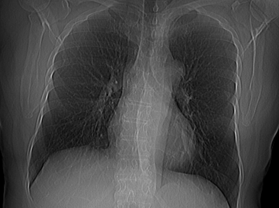
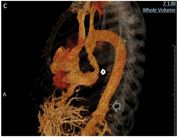
A 59-year-old Caucasian female, with no history of disease, was admitted to our emergency department with acute unexplained dyspnea and palpitation. On admission, physical examination revealed a dyspneic patient without any other findings. The family history was negative for remarkable diseases or congenital abnormalities. Electrocardiogram showed a regular rhythm, with sinus tachycardia. Blood pressure of the patient was normal. The room air oxygen saturation was 89%, and arterial blood gas analysis revealed hypoxemia with an elevated alveolo-arterial oxygen gradient. All blood tests, except D-dimer, were normal. Chest X-ray revealed superior mediastinum widening and double shadow of thoracic aorta (Figure 1), but no other remarkable sign. A transthoracic echocardiogram showed normal left ventricle function with a patent foramen ovale, and minor degree of tricuspid and mitral valve regurgitation. In order to exclude the diagnosis of pulmonary embolism and to investigate and evaluate the others suspected, combined abnormalities, a CT scan of chest and abdomen was performed. A persistent left SVC was revealed, connected with the right superior vena cava (SVC) by a bridging vein, which was crossing anterior to the aortic arch (Figure 2). The left SVC was running lateral to the aortic arch (Figure 3) and through the coronary sinus to the right atrium (Figure 4). Right lung was bilobed. A dilated hemiazygos vein was located posterior to the descending aorta and was draining to the left SVC. Left inferior vena cava (IVC) with hemiazygos continuation was also revealed. There was total absence of azygos vein. Hepatic veins were drained to right atrium. Portal vein was placed anterior to the midline gallbladder. The origin of left hepatic artery was the superior mesenteric artery. The liver was also midline and symmetric. Pancreas was truncated. The lobulated spleen, the multiple small satellite accessory spleens and the stomach were located at the left. Left-sided colon and right-sided small bowels indicated intestinal malrotation (Figure 5). The diagnosis was consistent with Situs Ambiguous with polysplenia syndrome, without additional findings. | ||||||
| ||||||
|
| ||||||
|
| ||||||
|
| ||||||
|
| ||||||
|
Discussion
| ||||||
|
Although situs ambiguous has been described in literature several times, we have found only few description of this entity presented in adults, and none completely consistent with our findings. This fact could be explained by the high mortality rates of the syndrome, which is consistent with the severity of the associated congenital heart disease [4]. However, patients with minor cardiac malformations may survive to adulthood [2]. Applegate et al. proposed an individualized approach on classification of heterotaxy syndrome based on the multiple possible combinations between anatomical variants [1]. They suggested that each case should be labeled heterotaxy syndrome followed by a description of the specific patient's anatomy [1]. Jacobs et al. also emphasize the importance of the exact description of cardiac anatomy and associated cardiac malformations, as well as the relationship and location of thoraco-abdominal organs [3]. There is a wide spectrum of abdominal abnormalities consist with situs ambiguous with polysplenia. Although associated with the presence of multiple discrete spleens in the majority of patients, also patients with a single, lobulated spleen or even normal spleen have been reported [2]. There are reports of young patients with altered splenic function [1] . The correlation between spleen and stomach location, can be interpreted by the fact that splenic tissue develops in the dorsal mesogastrium [2]. Often exists a bridging midline liver, but also right-sided and left-sided (rarely) liver has been reported [2]. The gallbladder is usually in the midline [2]. The pancreas can be truncated. Only the pancreatic head located to the right of midline or midline, accompanied or not, with a small portion of the pancreatic body is often present [2]. The stomach can be right-sided or left-sided [2]. Bowel's rotation abnormalities are usually observed [1]. Thoracic findings consist in bilobed lungs with hyparterial bronchi, levocardia more often than dextrocardia and congenital heart diseases, that may vary from minor or even absent, to severe, life-threatening ones [1] [2]. Among venous malformation, the congenital interruption of the IVC is the most common finding with azygos or hemiazygos continuation [1]. In these cases a short intrahepatic segment of inferior vena cava is present [1]. In addition, in our case a few more venous variations were observed. A left IVC, the embryological substratum of it resides in the persistence of the left supracardinal vein instead of the right one [5]. Total absence of azygos vein. A very rare anomaly, that arises when the right segment of the vein fails to develop [6]. A double SVC. It can be explained by persistence of right cardinal vein, as well as, by failure of left cardinal vein to regress [6]. The left-sided SVC typically is draining into the coronary sinus via the vein of Marshall [6]. Multiple modes of inheritance are proposed for heterotaxy syndrome, including autosomal dominant, autosomal recessive, and X-linked recessive mode of inheritance [1]. But a careful genetic study supports a multifactorial inheritance [7]. Abdominal organs and gastrointestinal tract misarrangements can create a confusing clinical picture, especially in the setting of abdominal diseases such as appendicitis, cholecystitis or volvulus, while the patient's pain and symptoms do not correlate with the expected locations of the affected organs [2]. Vascular anatomical variations can cause confusion during imaging reporting or even complications in the course of an invasive procedure. Venous abnormalities can be misinterpreted, on chest or abdominal X-ray films, as a mediastinal or retroperitoneal neoplasm, or lymphadenopathy [5]. Central venous cannulation may result in unusual catheter locations [6]. The persistent left SVC can disrupt manipulation of a cardiac venous catheter in or through the coronary sinus, which can result in hypotension, angina or cardiac arrest [8]. Additionally, the presence of this vessel has also been related to a higher risk of arrhythmias; most commonly atrial fibrillation [6]. The outcome of cannulation for cardiopulmonary bypass could lead to ineffective retrograde cardioplegia [6]. Unexpectedly, located vascular branches can cause life-threatening intraoperative hemorrhage due to vessels damage, during surgeries [6]. | ||||||
|
Conclusion
| ||||||
|
As the developmental abnormalities do not always cause clinical symptoms and as the use of imaging increases, situs abnormalities is likely to be detected with higher frequency in the future. So with great attention, should be considered their significance and their potential consequences, as the identification of high risk patients for congenital heart malformations, intestinal volvulus, atypical presentations of abdominal diseases or immune deficiency will improve their care. Careful analysis and understanding is mandatory, as these anatomical missarrangements can also cause confusion in differential diagnosis and severe clinical implication during invasive procedures. | ||||||
|
References
| ||||||
| ||||||
|
Suggested Reading
| ||||||
| ||||||
|
[HTML Abstract]
[PDF Full Text]
|
|
Author Contributions
Niki Lama – Substantial contributions to conception and design, Acquisition of data, Analysis and interpretation of data, Drafting the article, Revising it critically for important intellectual content, Final approval of the version to be published Petros Maniatis – Substantial contributions to conception and design, Acquisition of data, Analysis and interpretation of data, Drafting the article, Final approval of the version to be published Dionisios Haralambos Antonatos – Substantial contributions to conception and design, Drafting the article, Final approval of the version to be published Dimitrios Fagkrezos – Analysis and interpretation of data, Drafting the article, Final approval of the version to be published Charikleia Triantopoulou – Substantial contributions to conception and design, Acquisition of data, Analysis and interpretation of data, Drafting the article, Revising it critically for important intellectual content, Final approval of the version to be published Ioannis Papailiou – Analysis and interpretation of data, Revising it critically for important intellectual content, Final approval of the version to be published |
|
Guarantor of submission
The corresponding author is the guarantor of submission. |
|
Source of support
None |
|
Conflict of interest
Authors declare no conflict of interest. |
|
Copyright
© 2015 Niki Lama et al. This article is distributed under the terms of Creative Commons Attribution License which permits unrestricted use, distribution and reproduction in any medium provided the original author(s) and original publisher are properly credited. Please see the copyright policy on the journal website for more information. |
|
|
|
About The Authors
| |||
| |||
| |||
| |||
| |||
| |||
| |||