 |

|
 |
|
Clinical Image
| ||||||
| 101 spots: Find the primary site | ||||||
| Geraldine Bera1, Gabriel Malouf2, Nathanaëlle Yeni3, Charlotte Lepoutre-Lussey4 | ||||||
|
1University Medical Assistante, Department of Nuclear Medicine, AP-HP Hospital Pitié-Salpétrière, 47-83 Bd de l'hôpital 75651 Paris Cedex 13 France and Departement of Biophysic UPMC Paris VI, 91 Bd de l'hôpital 75651 Paris Cedex 13 France.
2Clinical University Hospital Asssitant, Department of Medical Oncology, AP-HP Hospital Pitié-Salpétrière, 47-83 Bd de l'hôpital 75651 Paris Cedex 13 France and Laboratoire de la Fondation AVEC and Institut Universitaire de Cancérologie, GRC5, UPMC, Paris, France. 3Contractual Hospital Practitioner, Department of Nuclear Medicine, AP-HP Hospital Pitié-Salpétrière, 47-83 Bd de l'hôpital 75651 Paris Cedex 13 France and Department of Biophysic UPMC Paris VI, LIB, INSERM UMR 678, 91 Bd de l'hôpital 75651 Paris Cedex 13 France. 4Practitioner Attached, Department of Biochemistry INSERM 0970, AP-HP European Hospital Georges-Pompidou, 56 rue Leblanc 75015 Paris, France ; Department of Nuclear Medicine, AP-HP Hospital Pitié-Salpétrière, 47-83 Bd de l'hôpital 75651 Paris Cedex 13 France. | ||||||
| ||||||
|
[HTML Abstract]
[PDF Full Text]
[Print This Article]
[Similar article in Pumed] [Similar article in Google Scholar] 
|


| How to cite this article |
| Bera G, Malouf G, Yeni N, Lepoutre-Lussey C. 101 spots: find the primary site. Int J Case Rep Images 2015;6(11):727–729. |
|
Case Report
|
|
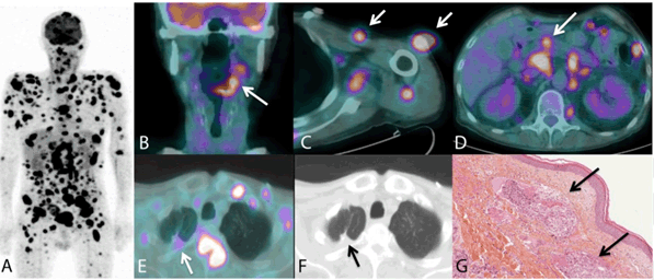
A 57-year-old male was presented to the emergency department with acute dysphagia and severe hypercalcaemia. He had a history of a locally advanced oral squamous cell carcinoma (OSCC) of the base of the tongue and a localised pleomorphic pulmonary sarcomatoid carcinoma (PSC) of the upper lobe of the right lung, treated five years and eighteen months ago, respectively. Clinical examination revealed a bleeding epiglottic lesion, cervical lymphadenopathies and scattered subcutaneous nodules. 18Fluorine-fluorodeoxyglucose positron emission tomography/computed tomography (18[F]-FDG PET/CT) scan highlighted intense whole-body disseminated uptake (Figure 1A), in particular from the oral cavity to pyriform sinus (Figure 1B), in the subcutaneous tissues (Figure 1C), on bone, pancreas (Figure 1D) and a moderate uptake on the right upper lung lobe probably corresponding to a pleomorphic PSC recurrence (Figure 1E, 1F). Thus these findings have suggested a distant metastatic relapse of the PSC associated with a loco-regional recurrence of OSCC. Surprisingly, histological analysis of subcutaneous biopsies (Figure 1G) concluded a well-differentiated SCC and immunostaining for thyroid transcription factor-1 (TTF-1) was negative. |
|
|
|
Discussion
|
|
Pulmonary sarcomatoid carcinoma is a rare, poorly differentiated subtype of non-small cell lung cancer accounting for 0.1–0.4% of all lung malignancies. They occur mostly in smoking males, at an average age of 60 years. With a high frequency of local recurrence and distant metastatio, they are responsible of an aggressive clinical course. Microscopically, this poor prognostic disease is the result of a biphasic proliferative malignant cells with both carcinoma and sarcomatoid components. Immunohistochemically, TTF-1, a specific marker of thyroid and pulmonary tumors such as adenocarcinoma and small cell carcinoma, is find to be positive in more than 50% of PSCs [1]. Head and neck squamous cell carcinoma (HNSCC), as far as they are concerned, preferentially metastasise to cervical lymph nodes. They have an uncommon hematogenous spread varying from 4.2–23.8%, up to 57% at autopsies on the lungs (80%), mediastinal nodes (34%), bone (31%) and liver (31%). Distant metastatic clearly affect the prognosis of HNSCCs and if not present at initial presentation it usually becomes apparent within two years [2]. Even so their clinicopathological predictive factors of occurrence are the site of the primary tumor (oropharynx, hypopharynx and larynx), multilevel nodal involvement in neck and primary tumor invasion into muscular, bone or cartilage ; metastatic screening by 18[F]-FDG PET/CT can detect them early, before the onset of medical complications [3]. Detection of occult disseminated disease without curative options allow avoiding all futile treatment, counseling patients about prognosis and optimizing their quality of life [4]. Cancer-related hypercalcemia is most common in patients with lung, breast, head and neck and kidney cancer. It could be related either to osteolysis of the severe bone metastases -that was probably the reason for this atypical patient- or a paraneoplastic syndrome. Having a strong correlation with the stage of the primary tumor and the development of recurrence or metastasis, hypercalcemia has an adverse impact on survival of OSCC. Its early recognition could help to recognize occult neoplasms leading to a proper therapeutic strategy and so prolong survival with a better quality of life [5]. |
|
Conclusion
|
|
This atypical case illustrates the utility of whole-body 18Fluorine-fluorodeoxyglucose positron emission tomography computed tomography (18[F]-FDG PET/CT) scan in the follow-up of head and neck squamous cell carcinoma (HNSCC) that can have a fatal issue due to distant metastatic and their resulting metabolic disorders such as hypercalcemia, more than their common local aggressive growth. Screening progression in these patients may help to anticipate the complications that are difficult to be managed. Keywords: Cancer, Distant metastasis, Pulmonary sarcomatoid carcinoma, Head and neck squamous cell carcinoma |
|
Acknowledgements
|
|
Professor Aurelie KAS MD. PhD., Department of Biophysic UPMC Paris VI, LIB, INSERM UMR 678, 91 Bd de l'hôpital 75651 Paris Cedex 13 France and Department of Nuclear Medicine, AP-HP Hospital Pitié-Salpétrière, 47-83 Bd de l'hôpital 75651 Paris Cedex 13 France. Pathology Department, AP-HP Hospital Pitié-Salpétrière, 47-83 Bd de l'hôpital 75651 Paris Cedex 13 France. Head and neck Oncology concertation meeting, AP-HP Hospital Pitié-Salpétrière, 47-83 Bd de l'hôpital 75651 Paris Cedex 13 France. |
|
References
|
|

|
[HTML Abstract]
[PDF Full Text]
|
|
Author Contributions
Geraldine Bera – Substantial contributions to conception and design, Acquisition of data, Analysis and interpretation of data, Drafting the article, Revising it critically for important intellectual content, Final approval of the version to be published Gabriel Malouf – Acquisition of data, Analysis and interpretation of data, Revising it critically for important intellectual content, Final approval of the version to be published Nathanaëlle Yeni – Acquisition of data, Analysis and interpretation of data, Revising it critically for important intellectual content, Final approval of the version to be published Charlotte Lepoutre-Lussey – Substantial contributions to conception and design, Revising it critically for important intellectual content, Final approval of the version to be published |
|
Guarantor of submission
The corresponding author is the guarantor of submission. |
|
Source of support
None |
|
Conflict of interest
Authors declare no conflict of interest. |
|
Copyright
© 2015 Geraldine Bera et al. This article is distributed under the terms of Creative Commons Attribution License which permits unrestricted use, distribution and reproduction in any medium provided the original author(s) and original publisher are properly credited. Please see the copyright policy on the journal website for more information. |
|
|