| |
|
|
|
Clinical Image
| ||||||
| A case of rhino-orbital-cerebral mucormycosis | ||||||
| Aishwarya Venkataraman1, Bridget Callaghan2 | ||||||
|
1Paediatric Registrar, International and Private Patients, Great Ormond Street Hospital NHS Trust, London, United Kingdom.
2Lead Consultant for International and Private Patients, Great Ormond Street Hospital NHS Trust, London, United Kingdom. | ||||||
| ||||||
|
[HTML Abstract]
[PDF Full Text]
[Print This Article]
[Similar article in Pumed] [Similar article in Google Scholar] 
|
| How to cite this article |
| Venkataraman A, Callaghan B. A case of rhinoorbital-cerebral mucormycosis. Int J Case Rep Images 2015;6(11):724–726. |
|
Case Report
| ||||||
|
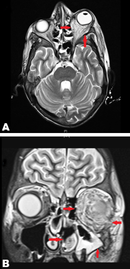
A 16-year-old, neutropenic boy on treatment for acute lymphoblastic leukemia developed acute onset fever and swelling of the left eye. The initial differential diagnoses were periorbital cellulitis and orbital cellulitis. Ophthalmological examination showed no eye movements, no pupillary reaction, no perception of light and a swollen retina. An urgent computed tomography (CT) scan of head with orbits was suggestive of orbital cellulitis and broad spectrum antibiotics (piperacillin/tazobactam and amikacin) were commenced. A detailed ENT examination on the same day revealed possible spores or hyphae along the nasal septum on the left side 2 cm from the nostril. Mucormycosis was suspected and he was started on antifungals (amphotericin B and posaconazole) in addition to the antibiotics. Magnetic resonance imaging (MRI) scan of the brain and orbits performed the next day, showed extensive infiltration consistent with infection of the left orbit, left maxillary antrum and ethmoidal sinus, extending to the right medial nasal region ( (Figure 1A), arrow and (Figure 1B), arrow). Subsequently, he underwent debridement and enucleation of the left eye with radical bilateral frontoethmoidectomy to salvage the right eye. Histology confirmed widespread necrotizing, angioinvasive and perineural fungal infection with involvement of sclera, choroid and dura. Culture grew Rhizopus microsporus, Staphylococcus aureus and Acinetobacter species. Despite optimum treatment, he succumbed to extensive central nervous system disease 40 days after initial presentation. | ||||||
| ||||||
|
Discussion
| ||||||
|
Mucormycosis is a rare, rapidly destructive fungal infection most commonly affecting immunocompromised and diabetic patients [1] [2] [3] [4]. Depending on the anatomical site involved, it can be broadly classified as rhino-orbital-cerebral, pulmonary, gastrointestinal, cutaneous or disseminated disease [1] [2]. The fungus is angioinvasive, causing severe thrombosis and tissue necrosis [1]. Underlying predisposing conditions are major risk factors for the disease [1] [2] [3][4]. In our case, chemotherapy-induced neutropenia (ANC of the patient remained <1.0x10⁹/l throughout) was one of the risk factors. Steroids, used in treatment of ALL, may also increase the risk of fungal infection [2][3]. The initial symptoms of rhino-orbital- cerebral mucormycosis is similar to either sinusitis or periorbital cellulitis. If untreated, infection can spread to the orbit from the ethmoid sinuses, resulting in loss of extraocular muscle function and proptosis. Progressive vision loss and ultimately blindness may result either from involvement of the optic nerve or from cavernous sinus thrombosis. Extensive central nervous system involvement can present with signs and symptoms of cerebral infarctions secondary to internal carotid artery and cavernous sinuses thrombosis [1]. Diagnosis of rhino-orbito-cerebral mucormycosis is challenging. Initial CT scan findings can be negative or have findings suggestive of sinusitis. MRI scans are more sensitive than CT scans for detecting disease beyond the sinuses [5]. Confirmatory diagnosis is made by histopathological examination and positive microbiology culture. Unfortunately, this is time consuming and Mucorales species often fail to grow in fungal culture [1]. Antifungal therapy alone is usually insufficient to control the rapidly spreading disease. It is important to treat or reverse the underlying cause and immunosuppressive medications should be dose reduced or stopped if possible [1] [2] [3]. Angioinvasion, thrombosis, and tissue necrosis result in poor penetration of anti-fungal agents to the site of infection. Surgical excision of the infected sinuses and appropriate debridement of the retro-orbital space is often required and can prevent the spread of infection into the eye, resulting in improved cure rates [1]. Unfortunately, mortality and morbidity remains high despite appropriate medical and surgical treatment [1] [6]. The patient described in the case report developed extensive cerebral and brain stem infarction due to the rapidly spreading disease despite prompt diagnosis and treatment. | ||||||
|
Conclusion
| ||||||
|
Rhino-orbital-cerebral mucormycosis is rare but potentially fatal in immunocompromised patients. Diagnosis is often difficult and surgical debridement, prompt use of antifungal agents and reversal of predisposing factors, if possible remains the mainstay of treatment. Despite optimum treatment, the mortality remains high; more than 50%. Newer tools are urgently needed to diagnose and treat mucormycosis. Keywords: Angio-invasive, Anti-fungal, Fungal infection, Mucormycosis, Posaconazole | ||||||
|
References
| ||||||
| ||||||
|
[HTML Abstract]
[PDF Full Text]
|
|
Author Contributions
Aishwarya Venkataraman – Substantial contributions to conception and design, Acquisition of data, Drafting the article, Revising it critically for important intellectual content, Final approval of the version to be published Bridget Callaghan – Substantial contributions to conception and design, Revising the article critically for important intellectual content, Final approval of the version to be published |
|
Guarantor of submission
The corresponding author is the guarantor of submission. |
|
Source of support
None |
|
Conflict of interest
Authors declare no conflict of interest. |
|
Copyright
© 2015 Aishwarya Venkataraman et al. This article is distributed under the terms of Creative Commons Attribution License which permits unrestricted use, distribution and reproduction in any medium provided the original author(s) and original publisher are properly credited. Please see the copyright policy on the journal website for more information. |
|
|