  |

|
 |
|
Case Report
| ||||||
| Adamantinoma of tibia: A case report | ||||||
| Rahadyan Magetsari1, Yudha M. Sakti1, Asa I. Z. Asikin1, Zikrina A. Lanodiyu1 Punto Dewo1 | ||||||
|
1Department of Orthopaedics and Traumatology, Dr Sardjito General Hospital/ Faculty of Medicine, Universitas Gadjah Mada, Yogyakarta, Indonesia.
| ||||||
| ||||||
|
[HTML Abstract]
[PDF Full Text]
[Print This Article]
[Similar article in Pumed] [Similar article in Google Scholar] 
|


| How to cite this article |
| Magetsari R, Sakti YM, Asikin AIZ, Lanodiyu ZA, Dewo P. Adamantinoma of tibia: A case report. Int J Case Rep Images 2015;6(10):627–631. |
|
Abstract
|
|
Introduction:
Adamantinoma is one of the rarest low-grade malignant bone tumors, representing 0.4% of them with only around 300 documented case of it.
Case Report: We reported a rare case of primary adamantinoma of the tibia in a 35-year-old Indonesian mongoloid female which recurred after excision of the primary tumor seven years prior to admission. Conclusion: Adamantinoma is hard to metastasize except in the case of repeated and unsatisfying removal procedure. Wide local excision with a substantial margin of normal bone can be applied if early diagnosis is successfully made. If there has been more than one recurrence or in large tumor with extension to the surrounding soft tissues, radical resection or amputation is advisable. After radical treatment, there is a high percentage of healing. The main factor of recurrence is incomplete resection. | |
|
Keywords:
Adamantinoma, Bone tumors, Primary adamantinoma, Tibia, Recurrence
| |
|
Introduction
| ||||||
|
Adamantinoma is one of the rarest low-grade malignant bone tumors, representing around 0.4% of them [1]. It was described initially by Fisher in 1913 and occurs most commonly between 10 and 50 years of age with slight male predominance. It was commonly found in the centerpart of long bone. However, some cases happened in other parts of the bone [2]. Around 90% of the cases there were reported previously was found in long tubular bones and 80% of it was found in the tibial mid shaft. Other long bones not uncommonly affected are the humerus, ulna, femur, fibula and radius. Early on it is confined to bone, furthermore there may be an extension inwards to the medullary canal or outwards beyond the periosteum. Pain is the most common clinical manifestation and local swelling is the common second clinical sign to appear. Distant metastases have been described to occur many years after the presentation of the primary lesion [2] [3] The incidence of recurrence is approximately 30% and those of metastases ranges between 10% and 20% [4]. We presented a case of adamantinoma of tibia with pneumothorax and suspected pulmonary metastasis with recurrence of the primary tumor after excision six years prior to admission. | ||||||
|
Case Report
| ||||||
|
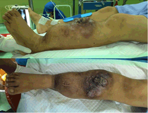
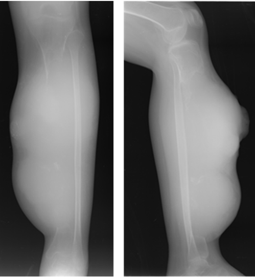
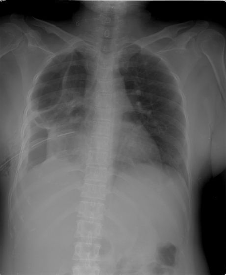
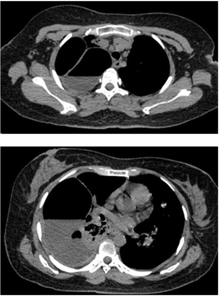
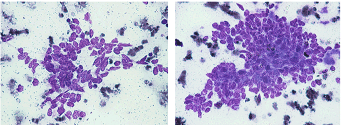
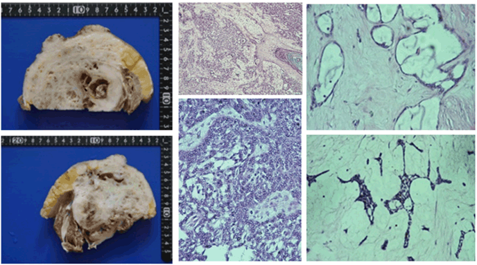
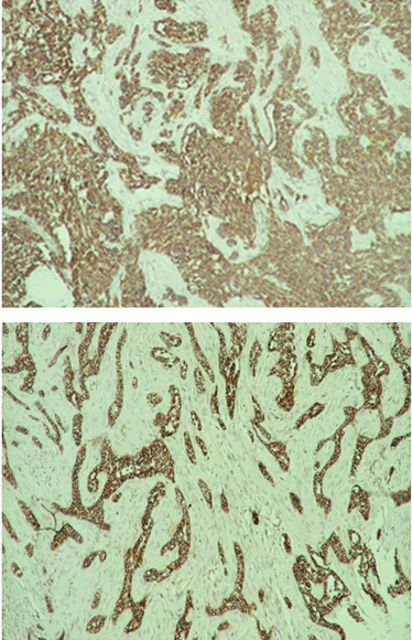
A 35-year-old Indonesian mongoloid female patient presented with a painful lump in her left leg. She had history of lump in her left leg since seven years prior to admission. Initially, the lump was only 1 cm in diameter. There was no history of trauma. The patient was initially treated by general surgeon, and had undergone excision of the lump. After the surgery, recurrence occurred and the lump developed gradually. Within seven years, the patient did not routinely visit hospital to have the condition of the lump checked. One week prior to admission, the patient complained about difficulty in breathing. She came to a primary hospital and was found to have a pleural effusion in her right lung and chest tube was inserted. She was then referred to our hospital. On the physical examination, a 10x10x5 cm hard tender mass was found in the left lower leg (Figure 1). Range of movement (ROM) in the left knee joint was full, but in the ankle joint, the ROM was limited. Laboratory results including tumor marker were unremarkable. Plain radiograph of the left leg showed lytic lesion in diaphysis and metaphysis of the left tibia with osteodestruction of tibia and extension to the surrounding soft tissue (Figure 2). A chest radiograph was performed (Figure 3) which showed bullae in the superior lobe of the right lung, pneumothorax, fibroatelectasis in the middle lobe of the right lung, and right pleural effusion. A chest MSCT showed hydropneumothorax and multiple bullae of the right lung and solitary nodule in the left lung suspected to be a distant metastasis from the primary tumor site (Figure 4). The patient was assessed with hydropneumothorax of the right lung caused by possible malignancy. A fine needle aspiration biopsy (FNAB) of the lesion in the left leg then performed with the result of clustered tumor cells within a group, with small cells with atypia, small amount of cytoplasm, spindle nucleus with hyperchromasia with background of lymphocyte and erythrocyte (Figure 5). A clinicopathological conference was then arranged with a recommendation of transfemoral amputation. A transfemoral amputation was then performed and the histopathological result showed a pattern of epithelial tumor with solid arrangement and infiltration to surrounding tissue, including to the upper dermis. The observation of the cells showed small to moderate cell sizes, small amount of cytoplasm, round to spindle nucleus with small amount of mitosis. The conclusion of the histopathological examination was islands of epithelial cells in a densely-populated stroma of spindle cells suggesting adamantinoma (Figure 6). The positive cytokeratin was found in the immunohistochemistry on the tumor cells and it was also relevant with the diagnosis of adamantinoma (Figure 7). | ||||||
| ||||||
|
| ||||||
| ||||||
| ||||||
|
| ||||||
|
| ||||||
| ||||||
|
Discussion
| ||||||
|
The symptoms initially present with nonspecific characteristics and its variety of the symptoms is related to location and extent of the disease. Pain is the most common clinical manifestation reported. It was in accordance to this case where the patient was presented with painful lump on her left lower leg. The onset of the clinical sign and symptom is insidious with slow and progressive characteristic. The patient usually tolerates symptoms for many years before seeking medical attention because of this characteristic of clinical manifestation as seen in this patient [5]. However, some of the patients present with swelling with or without pain as local swelling being the second most common symptom to appear. In addition, this case also showed that involvement of the anterior tibial surface can produce bowing of the tibia [6]. Based on radiographic assessment, adamantinoma is an osteolytic type with mono or multi-loculated lesions. In accordance to its benign characteristics, it is well circumscribed with septa and a peripheral condensation may appear. It is usually appear in the diaphysis or metaphysis of the anterior tibial. The lesion usually grows intracortical and it may spread longitudinally. However, it does not rule out the possibility of cortex destruction and marrow cavity invasion of the tumor [7]. The MSCT examination could reveal the characteristics of the lesion better than plain radiograph and can also detect another tumor site invisible on plain radiographs. The differential diagnosis based on plain radiograph examination including fibrous dysplasia and osteofibrous dysplasia [2]. . The patient in this case had tumor in her left leg which develops progressively after the first excision seven years ago. This might be due to inadequate attempts of removal where incomplete resection may result in higher recurrence rate up to 30% in the period of 85 months. Therefore, adamantinoma is suggested to be treated with wide surgical excision and reconstruction or amputation since its likelihood of healing is high subsequent to radical treatment [2] [3]. If early diagnosis is successfully made, wide local excision with a substantial margin of normal bone is still acceptable [8]. However, if there has been more than one recurrence or in large tumor with extension to the surrounding soft tissues, radical resection or amputation is advisable as shown in this patient where knee amputation was performed [9]. Adamantinoma is a low-grade malignant tumor of epithelial origin which metastasizes late. This tumor is insensitive to radiation and has capabilities of metastasis, especially to the lung. [8]. Metastases including in lung or lymph nodes are rare and can occur in 10–20% of patients. Pulmonary metastases are more common that regional nodal metastases from this tumor. It is not uncommon to develop distant metastases even up to 10 years after detection of the primary [3] [5]. This was in accordance with this case, where the main reason to seek medical management was due to difficulty in breathing and it was correlated with repeated local recurrences that might be due to inadequate primary excision of the tumor. However, it is difficult to evaluate its true metastatic potential due to several reasons such as long term follow-up, low number of the case, and slow growing nature of the tumor [5]. Several reports in literature have described metastasectomy for pulmonary lesions in both curative and palliative settings with good results. Mean survival of patients with metastatic disease is reported to be 12 years. There appears to be no definitive role for radiotherapy or chemotherapy [10]. Due to its excellent prognosis, it is crucial to diagnose this rare bone tumor in the early stage. It can be achieved by histologic examination, where this tumor can be distinguished easily. However, not only this tumor is rare, the heterogeneity of the tumor presentation may lead to confusion in some cases. On the histologic examination with inadequate sample taking, epithelial component may be seen only focally in the differentiated adamantinoma case, therefore in some cases, extensive sampling of the tumor is mandatory [11]. The challenge in making diagnosis and preparing proper management emphasizes that clinicopathological conference among orthopedic surgeon, pathologist, and radiologist was very important to ensure the patient get the best treatment available in musculoskeletal tumor cases. At the moment where the diagnosis is certain, resection with wide surgical margins or amputation can be applied to the patient.. | ||||||
|
Conclusion
| ||||||
|
We reported a rare case of recurrent tibial adamantinoma of the left lower leg with pulmonary metastases managed by knee amputation. Appropriate diagnosis and treatment plan through clinicopathological conference is mandatory to ensure the patient receive the best management in musculoskeletal tumor cases. | ||||||
|
References
| ||||||
| ||||||

|
[HTML Abstract]
[PDF Full Text]
|
|
Author Contributions
Rahadyan Magetsari – Substantial contributions to conception and design, Acquisition of data, Analysis and interpretation of data, Revising it critically for important intellectual content, Final approval of the version to be published Yudha M. Sakti – Acquisition of data, Analysis and interpretation of data, Revising it critically for important intellectual content, Final approval of the version to be published Asa IZ Asikin – Acquisition of data, Drafting the article, Final approval of the version to be published Zikrina A. Lanodiyu – Substantial contributions to conception and design, Drafting the article, Final approval of the version to be published Punto Dewo – Substantial contributions to conception and design, Analysis and interpretation of data, Drafting the article, Revising it critically for important intellectual content, Final approval of the version to be published |
|
Guarantor of submission
The corresponding author is the guarantor of submission. |
|
Source of support
None |
|
Conflict of interest
Authors declare no conflict of interest. |
|
Copyright
© 2015 Rahadyan Magetsari et al. This article is distributed under the terms of Creative Commons Attribution License which permits unrestricted use, distribution and reproduction in any medium provided the original author(s) and original publisher are properly credited. Please see the copyright policy on the journal website for more information. |
|
|